Reading...
![]()
Play button
![]()
Play button
![]()
Use LEFT and RIGHT arrow keys to navigate between flashcards;
Use UP and DOWN arrow keys to flip the card;
H to show hint;
A reads text to speech;
7 Cards in this Set
- Front
- Back
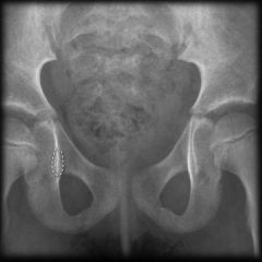
Which of the following is true regarding the structure outlined in Figure A? Topic Review Topic
FIGURES? |
In normal hips, all children usually have this radiographic figure by 18 months of age
The structure outlined in Figure A is the acetabular teardrop and it is comprised of the quadrilateral surface and cotyloid fossa. In normal hips, all children have a teardrop figure by age 18 months of age. |
|
|
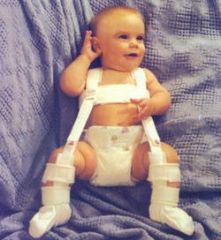
A 6-week-old female infant is referred to your practice for concerns of developmental dysplasia of the hip. On physical exam, you note a positive Ortolani test on the left side. Pavlik harness treatment is initiated. Which of the following imaging modalities should be utilized at the two week follow-up visit?
|
Ultrasound (US)
Initial ultrasound is performed to confirm reduction of the hip in question (generally after 1 or 2 weeks) followed by repeat ultrasound 6 weeks later. Ultrasound is necessary to avoid leaving an infant in a harness with an unreduced hip which can erode the acetabulum. |
|
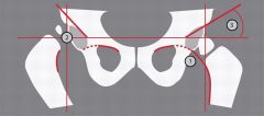
Which of the following best describes the radiographic measurement labeled #1 on Figure A. Topic Review Topic
FIGURES: A |
Radiographic line #1 on Figure A is consistent with Shenton's line.
Developmental dysplasia of the hip (DDH) refers to the the continuum of abnormalities involving the growing hip, (ranging from subluxation to dislocation of the hip joint). Shenton’s line is a projected arc from the inferior border of the femoral neck. Displacement of the femoral head or severe external rotation of the hip will result in a break in the continuity of Shenton’s line. Hilgenreiner’s line is a line drawn horizontally through each triradiate cartilage of the pelvis Perkins’ line is drawn perpendicular to Hilgenreiner’s line at the lateral edge of the acetabulum. The femoral head should lie within the inferomedial quadrant formed by Hilgenreiner’s and Perkins’ lines. |
|
|
A 15-year-old soccer player complains of bilateral hip pain. The pain is worse with activity and she notices that she has fatigue and pain that extends to the thighs and knees following a soccer match. She is nontender at the pubis symphysis and has no pain with resisted abdominal crunches. She has no pain with adduction of the hip. Hip flexion and rotation is normal. Which of the following is the next most appropriate step in treatment?
|
Peri-acetabular osteotomy (Ganz)
The peri-acetabular osteotomy (Ganz) is a reconstructive osteotomy for DDH patients with a closed triradiate cartilage. It allows for a large degree of three-dimensional correction because the cuts are close to the acetabulum as well as preserves the abductor muscles and allows for inspection of the joint. |
|
Which of the following concepts regarding pediatric hips is true?
|
The ossific nucleus of the proximal femur is visible on radiographs by 6 months of age in most children.
The proximal femoral physis and greater trochanteric apophysis develop from the same cartilage physis in the infant which undergoes apoptotic division in the child. The distal femoral physis (not proximal) grows at a rate of 9 mm per year. The normal infant femoral anteversion is between 30-40 degrees. SCFE typically occurs through the zone of hypertrophy, not the zone of proliferation. They recommond detection with ultrasound because of the delayed femoral head ossification (~5 months) and discuss the cost ineffectiveness of routine screening of all newborns. |
|
Failure to achieve reduction of a dislocated hip in an otherwise healthy 6 month old infant following 4 weeks in a Pavlik harness with the hips flexed to 90 degrees and abduction of 50 degrees is best treated with which of the following?
|
Closed reduction with hip arthrogram, adductor tenotomy if necessary, and hip spica casting
A 6-month-old who fails Pavlik harness treatment is best treated with closed versus open reduction of the hip and spica casting. Continued harness treatment can be detrimental as there is risk of posterior acetabular erosion. Osteotomies are not necessary to achieve reduction in a patient of this age cohort. if the hip does not become reduced within 2 weeks of treatment and recommends transitioning to alternative treatment spica cast |
|
|
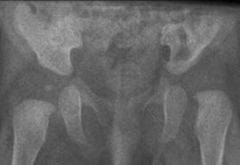
A 2-week-old infant girl is referred for a hip clunk noticed by the pediatrician. She was the product of a normal spontaneous vaginal delivery and is otherwise healthy. Examination demonstrates a right hip Ortolani sign. A coronal ultrasound is shown in figure A. What is the most appropriate next step in treatment?
|
Pavlik harness application
This patient has a right hip dislocation (DDH), as demonstrated by the positive Ortolani sign. Pavlik harness application is indicated for treatment. If the hip does not stay reduced within a few weeks, the next option is an arthrogram under anesthesia, closed reduction, and spica casting. Open reduction and casting is reserved for when closed reduction has failed. Acetabular osteotomy and femoral shortening are procedures used for children with DDH typically older than 1.5 years. |