![]()
![]()
![]()
Use LEFT and RIGHT arrow keys to navigate between flashcards;
Use UP and DOWN arrow keys to flip the card;
H to show hint;
A reads text to speech;
21 Cards in this Set
- Front
- Back
Abnormality? |
there is a thrombus in the inferior vena cava (IVC) |
|
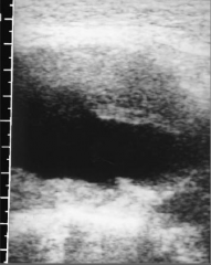
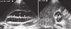
what does this longitudinal image demonstrate? |
this is a large abdominal aortic aneurysm with thrombus along the anterior and posterior borders. the lumen of the vessel is anechoic |
|
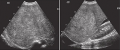
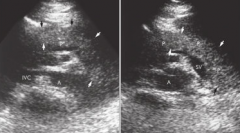
how can the sonographer determine that the inferior vena cava is dilated? |
if the inferior vena cava measures greater than 2.0 cm and does not show collapse with expiration, it is enlarged |
|
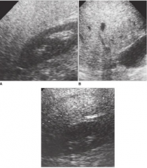
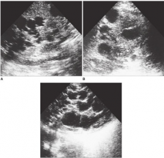
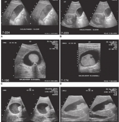
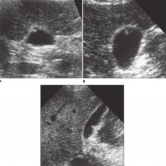
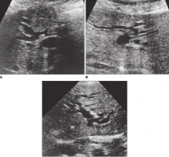
what liver disease is present in these images? |
these images show fatty infiltration of different grades
|
|
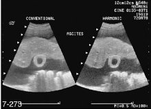
describe the abnormality in this image. |
image shows a shrunken liver with a coarse texture, consistent with cirrhosis. ascites surround the liver. the gallbladder wall is thickened, a false positive in patients w/ ascites; acute cholecysititis may not be ruled out by this finding alone |
|
what liver abnormality is demonstrated? what other area should the sonographer investigate? |
abnormality is polycystic disease
the sonographer should also investigate the kidneys |
|
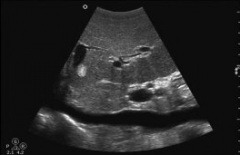
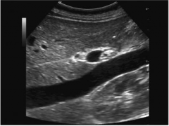
lesion seen in the right upper quadrant |
cavernous hemanigoma
a well-defined irregular lesion is seen in the dome of the right lobe of the liver .
differential considerations include metastasis, hepatoma (HCC), adenoma, and focal nodular hyperplasia |
|
a patient with a history of cirrhosis shows evidence of hepatomegaly. what are the possible sonographic findings? |
a large heterogenous in the right lobe of the liver extends from the dome of the liver, nearly filling the right lobe. this most likely represents a heptocellular carcinoma |
|
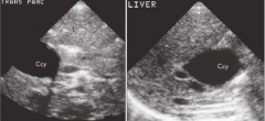
what is shown here?
what maneuvers may be performed to be sure the ____ in the gallbladder is not a tumor |
multiple patterns of sludge
sludge
change the patients position to see if the sludge moves. movement of sludge can be slow.
|
|
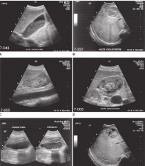
what is shown here?
describe clinical signs and sonographic signs |
acute cholecystitis
pain in the upper quadrant and fever are clinical signs. sono findings included enlarged gallbladder, positive murphys sign, thick gallbladder wall w/ irregularities. |
|
what anechoic structure(s) surround the gallbladder that may lead to confusion during examination? |
the duodenum may lie in the area of the gallbladder. if it is filled with fluid, this may lead to confusion and identification of the gallbladder. |
|
scan over right upper quadrant.
Representative of what disease |
choledochal cyst |
|
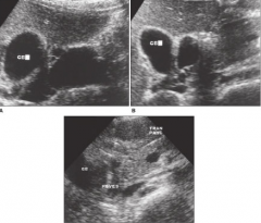
describe what arrows are pointing to in this image of the gallbladder? |
multiple stones are "floating" within the gallbladder with sludge along their posterior border |
|
describe the sonographic findings in these images |
cholesterol polyps
small, smooth wall projections seen to arise from the gallbladder wall. Multiple, do not shadow, or remain fixed to the wall with changes in patient position |
|
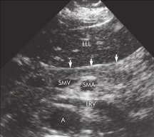
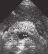
to which vascular structure are the arrows pointing in this image? |
a small segment of the superior mesenteric vein (arrows) is seen along the posterior border of the neck of the pancreas |
|
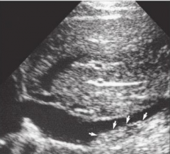
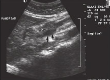
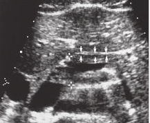
identify whether this image is transverse or longitudinal and what the arrows are pointing to |
the image is transverse, and the arrows are pointing to the pancreas |
|
identify the anatomic structure that the arrows are pointing to |
the collapsed wall of the stomach (arrows) may be seen as two parallel lines anterior to the body of the pancreas |
|
a 45-year-old male presents with midepigastric pain, elevated amylase and lipase levels, and tenderness.
identify the sono. findings |
sonographic findings consist of an enlarged edematous pancreas - pancreatitis |
|
patient with acute pancreatitis presents with continued pain.
describe sono. findings |
sonographic findings conisist of pancreatitis with hemorrhage. the gland is enlarged and echogenic secondary to freshly clotted blood |
|
56 yr old male with a 1-week of jaundice and pain has reported a 3-month history of nausea, vomiting, weight loss, and diarrhea.
What are the sonographic findings |
sonographic findings consist of adenocarcinoma of the pancreas with a dilated common bile duct. the gallbladder is dilated |
|
60 yr old female presents with a history of cholecystectomy several years ago. known to have had previoud hepatic calculi and now presents right upper quadrant pain.
What questions should be asked? describe sonographic findings |
how long has the patient experienced pain? where does the pain occur? does position change help? has the patient noticed yellow coloration in the whites of the eyes or skin?
there is a dilation of the intrahepatic ducts. carefully evaluate the liver and pancreas area for possible mass. |