Reading...
![]()
Play button
![]()
Play button
![]()
Use LEFT and RIGHT arrow keys to navigate between flashcards;
Use UP and DOWN arrow keys to flip the card;
H to show hint;
A reads text to speech;
66 Cards in this Set
- Front
- Back
|
What is the peritoneum?
|
Lines the peritoneal cavity. It is formed by a single layer of squamous epithelium on a thin layer of connective tissue. Consists of two continuous portions, the parietal peritoneum and the visceral peritoneum.
|
|
|
What is the parietal peritoneum?
|
Lines the abdominal walls.
|
|
|
What is the visceral peritoneum?
|
Covers the abdominal organs.
|
|
|
What moistens the peritoneal cavity?
|
Peritoneal (serous) fluid.
|
|
|
What is the peritoneal cavity?
|
B/t the visceral and parietal peritoneum, is a potential space. Completely empty, except for peritoneal fluid. Air can be admitted during surgery or when excess fluid (ascites) or pus accumulates.
|
|
|
What are the most important functions of the peritoneum?
|
1. produce peritoneal fluid
2. minimize friction (provides a slippery surface that permits free movement of abdominal viscera) 3. resist infection (tends to wall off infections) |
|
|
What is the less important function of the peritoneum?
|
Storage of fat, especially in the greater omentum.
|
|
|
What are the divisions of the peritoneal cavity?
|
Two continuous divisions
-lesser sac (omental bursa) -greater sac |
|
|
What is the lesser sac?
|
Located posterior to the lesser momentum, stomach, and caudate lobe of liver.
|
|
|
Is the lesser sac completely closed off from the greater sac?
|
It is closed off from the major peritoneal cavity (greater sac) except for the epiploic foramen.
|
|
|
What is the greater sac?
|
The major peritoneal cavity.
|
|
|
What borders the epiploic foramen?
|
-hepatoduodenal ligament (anteriorly)
-reflection of the peritoneum from the liver (superiorly) -duodenum (inferiorly) -parietal peritoneum covering the inferior vena cava (posteriorly) |
|
|
What is the mesentery?
|
A double-layered membrane of peritoneum which provides a channel through which vessels, nerves and lymphatics travel to reach various organs.
|
|
|
Where is the mesentery attached?
|
To the posterior abdominal wall where the blood and nerve supply to the abdominal viscera originates. Associated with the small intestine.
|
|
|
What is the omentum?
|
A mesentery extending from the stomach to adjacent organs. Two types:
-lesser omentum -greater omentum |
|
|
What is the lesser omentum?
|
It joins the lesser curvature of the stomach and the first part of the duodenum with the liver.
|
|
|
What is the greater omentum?
|
It joins the greater curvature of the stomach with the transverse colon.
|
|
|
What are the peritoneal recesses?
|
Blind pouches or tubular spaces opening into the peritoneal cavity.
|
|
|
What are the boundaries of the peritoneal cavity?
|
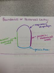
Anterior - anterior abdominal wall
Posterior - posterior abdominal wall Superior - respiratory diaphragm Inferior - pelvic diaphragm |
|
|
What is the esophagus?
|
Passes through the esophageal hiatus in the respiratory diaphragm to enter the abdomen. Has inferior esophageal sphincter.
|
|
|
What is the inferior esophageal sphincter?
|
NOT an anatomical sphincter!!! Just functional.
The respiratory diaphragm effectively closes the inferior portion of the esophagus |
|
|
What is the stomach?
|
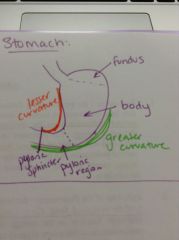
2-4 liters in volume, size varies depending on the person. Made up of 3 main parts:
-fundus -body -pylorus |
|
|
What is the fundus of the stomach?
|
It is the part of the stomach superior to the cardiac orifice (junction with esophagus) at the level of the 5th rib. It rests against the left side of the diaphragm.
|
|
|
What is the body of the stomach?
|
Just the portion between the fundus and pylorus.
|
|
|
What is the pylorus of the stomach? Its features?
|
Between the body and the duodenum. Has three main features:
-pyloric antrum (wide portion) -pyloric canal (narrow, distal portion) -pyloric sphincter |
|
|
What is the function of the pyloric sphincter?
|
Regulates the flow of food into the duodenum. Normally closed.
|
|
|
What is the greater curvature of the stomach?
|

Seen on the inferior border, is 4-5 times greater in length than the lesser curvature. The greater omentum attaches here.
|
|
|
What is the lesser curvature of the stomach?
|

Seen on the superior border, the lesser omentum attaches here.
|
|
|
What is the spleen?
|
It is NOT part of the GI tract, is a lymphatic organ in the LUQ. Is the most fragile organ in the abdomen, but you can live w/o it.
|
|
|
What happens to a person w/o a spleen?
|
They may become more susceptible to bacterial infections b/c the spleen is part of the immune system.
|
|
|
What is the gastrolienal ligament?
|
Connects the hilum of the spleen with the greater curvature of the stomach.
|
|
|
What is the lienorenal ligament?
|
Connects the hilum of the spleen to the left kidney.
|
|
|
What are the general characteristics of the duodenum?
|
-shaped like a horseshoe, molded around pancreas
-joins the pylorus to the jejunum -is the most fixed part of the small intestine, the last 9" is retroperitoneal (only 10" in length) -connects with the common bile duct and the pancreatic ducts -MOST IMPORTANT part of small intestine |
|
|
How many parts does the duodenum have?
|
Four. The first is completely surrounded by peritoneum, the remaining 3 parts are retroperitoneal (attached to posterior abdominal wall)
|
|
|
What is the first part of the duodenum?
|
Attached to stomach, completely surrounded by peritoneum. Only 1" in length, is the most mobile part of SI. Courses superior, posterior and to the right (up and to the right)
|
|
|
What is the second part of the duodenum?
|
3-4" in length. Courses parallel and to the right of the inferior vena cava (vertical). Contains the:
-hepatopancreatic ampulla of Vater -major duodenal papilla -sphincter of Oddi |
|
|
What is the hepatopancreatic ampulla of Vater?
|
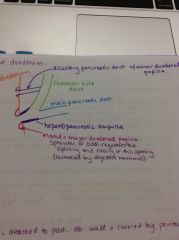
It is part of the 2nd part of the duodenum, formed by the union of the common bile duct and the pancreatic duct.
|
|
|
Where do the main pancreatic duct and accessory pancreatic ducts open into the duodenum?
|
Main - major duodenal papilla
Accessory - minor duodenal papilla |
|
|
What is the third part of the duodenum?
|
4" in length, courses from right to left horizontally
|
|
|
What is the fourth part of the duodenum?
|
Only 1" in length, ascend to the left side of the aorta. Contains the:
-duodenojejunal junction -ligament of Treitz |
|
|
What is the duodenojejunal junction?
|
(Or flexure), is a sharp bend that occurs where the duodenum meets the jejunum
|
|
|
What is the ligament of Treitz?
|
A suspensory muscle that attaches the duodenum and jejunum to the posterior abdominal wall. Widens this portion of the duodenum.
|
|
|
What is the clinical significance of the ligament of Treitz?
|
If in a high impact car accident, the inertia will be exerted through this ligament and can lead to tears and internal bleeding.
|
|
|
What is the pancreas?
|
Located just posterior to the lesser sac, is retroperitoneal. Functions as an endocrine and exocrine gland.
|
|
|
What are the parts of the pancreas?
|
Head - in concavity of duodenum
Neck - joins the head and the body, overlies the superior mesenteric a. Body Tail - b/t the two layers of lienorenal ligament, tip is related to hilum of spleen |
|
|
What is the clinical significance of the tail of the pancreas?
|
When removing the spleen during surgery, it has to be carefully avoided to not damage the pancreas.
|
|
|
What are the ducts of the pancreas?
|
-main pancreatic duct
-accessory pancreatic duct -hepatopancreatic ampulla |
|
|
What is the main pancreatic duct?
|
It begins in the tail and is usually joined by the accessory duct in the head, contains the sphincter of the main pancreatic duct, which prevents bile from entering the pancreas.
|
|
|
What is the accessory pancreatic duct?
|
Inconsistent feature, only in some people. If it does not join the main duct, then it has a separate opening in the duodenum located at the minor papilla.
|
|
|
What is the hepatopancreatic ampulla?
|
Formed by the union of the common bile duct and the main pancreatic duct. Associated with the sphincter of Oddi and the major duodenal papilla.
|
|
|
What is the location and function of the liver?
|
Located in URQ or epigastric and R hypochondriac regions.
Produces bile, processes nutrients absorbed by the GI tract, detoxifies xenobiotic chemicals, phagocytosis of worn out blood cells. |
|
|
What are the general features of the liver?
|
-diaphragmatic surface
-visceral surface -porta hepatis -L sagittal fissure -R sagittal fissure -R and L lobes |
|
|
What is the diaphragmatic surface?
|
Conforms to the concavity of the diaphragm. Covered by peritoneum except where in direct contact with the diaphragm (bare area of liver). Inferior vena cava directly touches this surface.
|
|
|
What is the porta hepatis?
|
"Window of liver". A transverse fissure in the visceral surface, contains the hepatic portal vein, hepatic proper artery, nerve plexus, hepatic (bile) ducts and lymphatic vessels.
|
|
|
What is contained within the left sagittal fissure?
|
Visceral surface. The ligamentum teres (the obliterated umbilical vein)
|
|
|
What is contained within the right sagittal fissure?
|
Visceral surface. Contains the gall bladder and inferior vena cava.
|
|
|
What is contained within the right lobe?
|
The quadrate and caudate lobes, which are separated by the porta hepatis. Is the larger lobe.
|
|
|
What is contained within the left lobe?
|
Separated from the R lobe to falciform ligament.
|
|
|
What are coronary ligaments?
|
A reflection of visceral peritoneum from the liver to the respiratory diaphragm, surrounding the bare area of the liver.
|
|
|
How does the lesser omentum relate to the liver?
|
-hepatogastric ligament
-hepatoduodenal ligament |
|
|
What is the hepatogastric ligament?
|
Passes from the porta hepatis of the liver to the lesser curvature of the stomach.
|
|
|
What is the hepatoduodenal ligament?
|
Passes from the porta hepatis of the liver to the first part of the duodenum, contains the proper hepatic a., hepatic portal v., common bile duct, lymphatic vessels and hepatic nerves.
|
|
|
What is the relation of hairy chests and cirrhosis of the liver?
|
Men who have hairy chests are less likely to have cirrhosis of the liver. Also, Jewish people are not susceptible to cirrhosis. People who live closer to the equator are more vulnerable to cirrhosis.
|
|
|
What is the biliary system pathway?
|
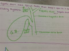
-microscopic bile canaliculi which collect bile from individual liver cells
-canaliculi merge to form interlobular bile ducts merge to form L and R hepatic ducts -R and L hepatic ducts merge to form the common hepatic -common hepatic duct merges with the cystic duct to form the common bile duct -common bile duct contains a sphincter which prevents pancreatic secretions from entering the biliary system |
|
|
What is the gallbladder?
|
Stores and concentrates bile. Shaped like a pear, size 1-2 ounces. Has peritoneal covering on posterior and inferior surfaces.
|
|
|
What are the parts of the gallbladder?
|
Fundus - projects below the inferior border of the liver
Body - contacts the visceral surface of liver Neck - narrow and tapered, "S-shaped", contains a spiral muscosal fold keeps the lumen open |