Reading...
![]()
Play button
![]()
Play button
![]()
Use LEFT and RIGHT arrow keys to navigate between flashcards;
Use UP and DOWN arrow keys to flip the card;
H to show hint;
A reads text to speech;
185 Cards in this Set
- Front
- Back
|
A detector operated in which mode detects individual photon interactions and processes each event in real time?
|
Pulse mode. These detectors typically permit the energy associated with each event to be determined. This enables advanced features such as energy discrimination (rejection of events that do not meet preset criteria). This is a feature that is essential for quantitative counting applications and is especially crucial for imaging applications since image contrast is strongly correlated with the amount of scatter (relative to primary unscattered photons) accepted by the imaging system during acquisition. One significant drawback to this mode of operation is that there is a finite time required for processing of each individual event during which the system is not available to count additional events. This phenomenon is referred to as dead time (both physical and electronic stages contribute to detector dead time though the electronic stage usually dominates).
|
|
|
The well-counter and scintillation camera are two examples of detectors operated in what mode?
|
Pulse mode.
|
|
|
Detectors operated in what mode integrate collected charge so that the detector output is a current with amplitude that is proportional to the average rate of energy deposition in the detector?
|
Current mode. Detectors operated in this mode are much less susceptible to deadtime and can be used in applications where there is a high flux of radiation, making them ideal for high countrate and exposure rate surveying applications.
|
|
|
Detectors operated in what mode permit only a cumulative estimate of energy deposited within the detector in a given time period?
|
Integrated (absorbed) dose mode. These kinds of detectors are most often associated with personnel dosimetry. Those personnel dosimeters which provide an electronic reading in real-time have on-board micro-circuitry and have an integrated physical and electronic stage as a result. However, others which store absorbed dose in the transducer until the transducer is “read out” later using a specialized reader, have separate physical and electronic stages. The Thermoluminescent Dosimeter (TLD) and Optically-Stimulated Luminescent Dosimeter (OSL Dosimeter) are both examples of this latter category of dosimeters.
|
|
|
How do you calculate the Intrinsic Efficiency of a detector?
|
# of photons detected or absorbed/# of photons incident. Intrinsic detection efficiency decreases with increasing photon energy for a given material. It also increases with increasing density and atomic number..
|
|
|
How do you calculate Geometric Efficiency of a detector?
|
# of photons incident on a detector/# of photons emitted from a source. In a simple configuration where a point source is positioned some distance from a detector, the geometric efficiency decreases with distance from the source because the number of photons incident on the detector decreases with distance in proportion to the square of the distance between the source and detector as predicted by the inverse square law. Or, more intuitively, as the detector is moved closer to the source the fraction of photons incident on the detector relative to the total number of emitted photons, increases.
|
|
|
How do you calculate the Total Efficiency of a detector?
|
Total = Intrinsic efficiency x Geometric efficiency. Since by definition the intrinsic and geometric efficiencies are relative efficiencies and as such are < 1, the total detection efficiency will also be < 1. Example: The total efficiency of a well counter detector can be determined using a calibrated radioactive source sample with a known activity. The efficiency is computed from the ratio of the recorded counts per minute (cpm) to the known activity in Becquerels or disintegrations per minute (dpm).
|
|
|
Photon counting detectors that process individual radiation interactions require a finite interval of time to process these events. This interval of time, t is referred to as what?
|
The system "deadtime" because during this interval the system is unresponsive (dead) to additional interactions.
|
|
|
For detectors that experience ____ deadtime, the period over which the detector is unresponsive is not impacted by a second interaction and when the system has completed processing of the 1st event it is again free to process additional events.
|
Non-paralyzable. Both of these kinds of detectors experience counting losses with increasing activity (and therefore input counting rate) but the paralyzable system experiences a large drop in sensitivity at higher counting rates as the system becomes essentially “paralyzed” and unable to effectively process additional incoming events.
|
|
|
In systems that experience ____ deadtime, the arrival of a second event extends the deadtime interval.
|
Paralyzable. Both of these kinds of detectors experience counting losses with increasing activity (and therefore input counting rate) but the paralyzable system experiences a large drop in sensitivity at higher counting rates as the system becomes essentially “paralyzed” and unable to effectively process additional incoming events. A scintillation camera is an example of a detector that experiences paralyzable deadtime at high countrates with degradation of image quality due to count losses and mis-positioning of events.
|
|
|
A scintillation camera is an example of a detector that experiences ____ deadtime at high countrates with degradation of image quality due to count losses and mis-positioning of events.
|
Paralyzable.
|
|
|
Regarding gas-filled detectors, when the ions are collected a current is generated and this current magnitude is directly related to the ____ of ion collection.
|
Rate. The average energy expended in the creation of an ion pair is a function of the type of radiation, its energy (E) and the atomic number (Z) and density (ρ) of gas in the chamber. It is important to note that not all energy deposited in the detector results in ionization, some of the energy is absorbed via excitation of gas molecules.
|
|
|
Regarding gas-filled detectors (specifically the cylindricalelectrode configuration), what are examples of direct vs indirect interactions?
|
Direct = Charged β- particles that penetrate the detector entrance window directly ionize gas molecules.
Indirect = X-rays or gamma rays more typically indirectly ionize gas molecules when secondary fast electrons (β- particles) are produced after an initial Compton or Photo-electric interaction in the walls of the detector surrounding the gas. This is because the low attenuation or absorption coefficient for x-ray or gamma rays in gases leads to low probability of interaction by comparison with charged particles. |
|
|
In the gas-filled detector, the number of PRIMARY ion pairs formed is proportional to the energy deposited within the detector transducer. However the number of PRIMARY ion pairs formed is not necessarily equal to the number of PRIMARY ion pairs collected because of what two processes?
|
1. Recombination (of PRIMARY ion pairs)
2. Secondary Ionizations |
|
|
Regarding the Operating Voltage and Performance of Gas-Filled detectors, when does recombination occur?
|
Recombination can occur when either of the primary ions formed after ionization of a gas molecule (the +ve ion or negative electron, e-), combines with an oppositely charged ion before being collected at the electrodes. Since the probability of recombination increases with the time that the ion pair spends in the vicinity of other ion pairs, recombination occurs when the applied electrode voltage is not sufficient to accelerate and collect the initial ion pair quickly. Recombination effects decrease collection efficiency.
|
|
|
Regarding the Operating Voltage and Performance of Gas-Filled detectors, when do secondary ionizations occur?
|
Secondary ionizations occur (and collection efficiency increases) whenever the applied voltage is high enough that primary ion pairs formed gain sufficient kinetic energy that they in turn induce additional ionizations, thereby amplifying the effects of the radiation interaction that resulted in the production of the initial ion pair.
|
|
|
What strongly determines the efficiency of ion-pair collection and therefore the response of the gas-filled detector>
|
The magnitude of applied voltage across the electrodes.
|
|
|
In the gas-filled detector response curve, where is the Recombination region?
|
The left-most portion of the graph. Ionization detectors are not operated in the Recombination Region due to very low efficiency and non-linear performance with slight voltage fluctuations.
|
|
|
In the gas-filled detector response curve, where is the Saturation region?
|
The right-most portion of the graph. Detectors are not operated in the Saturation Region because the very high operating voltages may cause spontaneous ionization of gas molecules or “discharge” that is unrelated to the presence or magnitude of ambient radiation and irreparably damage the detector can result.
|
|
|
On the gas-filled detector response curve, from left to right, what are the three principle operating regions?
|

|
|
|
Ionization detectors have a stable response across a relatively wide voltage range (See Region I) and a relatively low sensitivity in comparison with other gas-filled detectors and therefore they are usually used in ____ mode.
|
Current.
|
|
|
What gas-filled detector would be useful in applications where accurate estimates of deposited energy (or exposure) are essential?
|
Ionization detectors.
|
|
|
What type of detector is the dose calibrator?
|
The dose calibrator, which is a gas-filled ionization detector with a well-shaped gas containment compartment, usually employs pressurized gas such as Argon (Ar: Z=18) for improved sensitivity to x-ray and gamma ray photon interactions.
|
|
|
Detectors operated in the Proportional Region experience a phenomenon known as ____ where the applied voltage is sufficiently high that the electrons being accelerated gain enough kinetic energy that they can initiate multiple secondary ionizations.
|
Gas amplification. These detectors are orders of magnitude more sensitive than ionization detectors, allowing them to be operated in pulse mode for gamma ray spectrometry applications. However, proportional counters are not routinely found in the clinical nuclear medicine department.
|
|
|
Detectors operated in the Geiger-Mueller region also experience the ____ effect, however the operating voltages in this region are so high that the number of secondary ion pairs formed reaches a saturation point.
|
Gas amplification. This produces a maximum number of ion pairs independent of the energy deposited in the detector during an interaction. Thus G-M detectors are extremely sensitive to radiation interactions but they are not useful for energy discrimination.
|
|
|
G-M detectors are operated in ____ mode and have a deadtime on the order of ____.
|
Pulse, 10-100 μsec. Because they are relatively sensitive in comparison to ionization detectors they are useful for low-level radioactive surveys but are not useful in very high radiation fields. They are also not useful for accurate exposure estimates, though a G-M detector calibrated to the energy of interest can be used for approximate estimates of exposure for radiation surveying purposes.
|
|
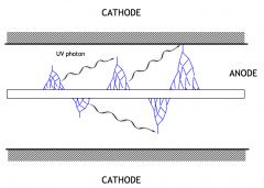
What does this diagram demonstrate?
|
Diagram showing the G-M gas amplification avalanche process. The accelerated electron induces additional excitations and ionizations. When excited electrons return to the ground state the UV photon produced can induce additional ionizations which can trigger additional cascades, propagating the effect until a saturation point is reached.
|
|
|
The mechanism of radiation interaction in solid state or semiconductor detectors is analogous to that of gas-filled detectors in that radiation interacting in the solid creates charge carriers but in this case because of the structure of solids, the charge carriers are in the form of ____ (in the conduction band) and ____ called “holes” (in the valence band)
|
electrons, electron vacancies
|
|
|
In solid state detectors, the energy gap separating the valence band from the conduction band, is referred to as the ____.
|
band gap energy. It is worth noting that the average energy expended to create an ion pair (W) is higher than the band gap or ionization energy because, as noted previously, the energy deposited in the detector yields both excitations and ionizations.
|
|
|
Which is more efficient, a solid state detector or a gas-filled ionization detector?
|
The efficiency of solid state detectors is an order of magnitude greater than gas-filled ionization detectors because 1. The density of solid state materials is a thousand fold greater than the density of gases and 2. the average energy expended to create an ion pair (W) is typically 3-5 eV which is much smaller than the approximately 34 eV expended to create an ion-pair in air.
|
|
|
Historically, solid state detectors used in nuclear counting applications were fabricated with Lithium-doped germanium and silicon, Ge(Li) and Si(Li), respectively. These detectors have largely been replaced by the high purity or intrinsic semiconductor, ____.
|
germanium detector (HPGe)
|
|
|
In Germanium or Silicone detectors, normally ambient room temperatures are sufficiently high that simple thermal conduction provides the energy needed to bridge the band gap energy threshold, resulting in ____ of germanium atoms
|
spontaneous ionization. Therefore, the HPGe detector must be cooled to very low temperatures using liquid nitrogen (or other sophisticated cooling systems) in order to avoid the significant electronic noise that would otherwise be associated with operation of the detector at room temperature. The HPGe detector is not routinely found in the clinical nuclear medicine setting. However, because it is useful in gamma ray spectroscopy applications, it is often used to evaluate radioisotopes for the presence of radioisotopic impurities.
|
|
|
The energy resolution achievable with a solid state is significantly better than that obtained using a scintillation detector, due to the ____ of the solid state detector.
|
high intrinsic efficiency
|
|
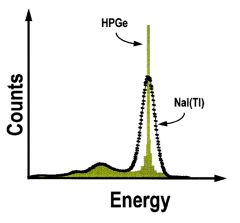
What does this graph demonstrate?
|
For High Purity Germanium (HPGe) shown here, the energy resolution is an order of magnitude smaller than that for NaI(Tl). In a HPGe detector, it takes approximately 3 eV to create an electron-hole pair. Therefore, approximately 333 electron-hole pairs will be created per 1 keV of energy deposited in the HPGe detector. By comparison, the number of photoelectrons produced at the cathode of a PMT in a typical NaI(Tl) scintillation detector per 1 keV of energy deposited in the crystal is < 10. [Note: the PMT gain yields an amplified signal but this does not change the underlying statistical fluctuation in measured photon energies which is determined by the number of photocathode electrons produced per unit of energy deposited in the crystal.]
|
|
|
The requirements for cooling associated with intrinsic (pure) semiconductors can be overcome by using compound semiconductor materials composed of high ____.
|
Z elements. Compound semiconductors can be used in detectors operated at room temperature because compound semiconductor band-gap energies are typically higher than those associated with intrinsic semiconductors.
|
|
|
Three examples of compound semiconductors encountered in imaging systems used in nuclear medicine applications are ____.
|
cadmium telluride (CdTe), cadmium zinc telluride (CdZnTe), and mercuric iodide (HgI2)
|
|
|
In solid state pixellated arrays, interactions in the CZT (CdZnTe) layer create ____ which are collected and read out using the application-specific integrated circuit (ASIC) at the bottom of the module.
|
charge carriers, Many such individual modules can be arranged to form a larger detector area more appropriate for clinical applications.
|
|
|
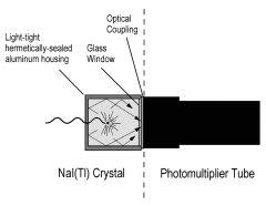
In scintillation detectors, the transducer material is a crystalline solid that has usually had trace amounts of ____ added during manufacturing.
|
impurity atoms
|
|
|
In scinitillation detectors, the doped scintillation crystals have the property of releasing ____ or ____ shortly after a radiation interaction in which a Compton or photoelectric interaction occurs in the crystal
|
visible, ultra-violet light
|
|
|
In scinitillation detectors, the doped scintillation crystals have the property of releasing visible or ultra-violet light shortly after a radiation interaction in which a ____ or ____ interaction occurs in the crystal.
|
Compton, photoelectric; In both cases the interaction produces a fast-moving electron that travels a short distance in the crystal, exciting additional electrons. These excited electrons migrate to the impurity sites called activation centers, releasing a burst of scintillation photons with an energy distribution that is characteristic of the scintillator.
|
|
|
In Thallium-doped sodium iodide or NaI(Tl) crystals, the scintillation crystal most commonly used in nuclear medicine scintillation detectors, the scintillation photons have an energy on the order of about ____ on average.
|
3 eV
|
|
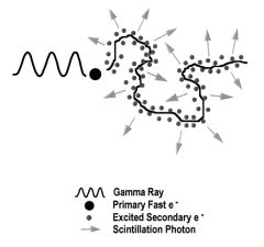
What does this graph depict?
|
Gamma ray interaction in a scintillation detector.
|
|
|
The NaI(Tl) crystal which is normally transparent has the property of being ____ which means that if the crystal is exposed to water or normal air containing trace amounts of humidity, the crystal will eventually lose its transparency and becomes hydrated or “yellowed”, significantly decreasing its efficiency.
|
hygroscopic;
|
|
|
NaI(Tl) crystals are encapsulated in an air-tight enclosure. Despite these precautions it is not unusual for a NaI(Tl) crystal to lose efficiency slowly over time as it loses transparency due to ____.
|
hydration
|
|
|
What are the five common scinitillation crystals?
|
NaI(Tl) – Thallium-activated sodium iodide (NaI:Tl)
BGO – Bismuth Germanate (Bi4Ge3O12) LSO – Lutetium Orthosilicate (Lu2SiO5) GSO – Gadolinium Orthosilicate (Gd2SiO5) YSO – Yttrium Orthosilicate (Y2SiO5) |
|
|
What is the only scinitllation crystal that is hygroscopic?
|
NaI(Tl)
|
|
|
The scintillation crystal characteristics strongly impact performance. Both density and effective atomic number have a direct impact on detection efficiency, as both increase, detection efficiency ____.
|
increases
|
|
|
What happens to the signal input as the relative scintillation light output increases?
|
The higher the relative scintillation light output, the better the characteristics of the signal input to the electronic stage and the detection system is therefore able to more accurately estimate the energy deposited in the detector.
|
|
|
In scintigraphy, energy resolution improves (Photopeak FWHM decreases) with ____ light output.
|
increasing
|
|
|
A detector employing a crystal with a ____ scintillation decay time will also be better able to maintain a separation between the light output produced by two events falling within a short interval of each other which is an important requirement if the electronic stage is to be able to process these events individually.
|
shorter
|
|
|
For a scintillation detector, overall system deadtime is comprised of ____ deadtime (within the crystal transducer) and ____ deadtime (within the electronic stage).
|
physical, electronic
|
|
|
A specialized amplification device called a ____ is coupled to the crystal in order to collect scintillation photos and convert them to a useful electronic signal.
|
photomultiplier or PMT
|
|
|
The outside of the crystal is painted with a reflective material to promote internal reflection of light so that a significant fraction (up to ____%) of the light produced within the crystal is collected at the PMT.
|
30
|
|
To maximize light transmission at the boundary between the crystal and PMT, an ____ is used to match the index of refraction of the crystal exit window and PMT entrance window.
|
optical gel or epoxy
|
|
|
The scintillation photons entering the PMT impact the ____ depositing their energy.
|
photocathode
|
|
|
For every five scintillation photons impacting the photocathode in a PMT, ____ electron is emitted.
|
one; That electron is then accelerated to the first of a series of shaped electrodes called dynodes, each of which is at a progressively higher voltage or potential. The accelerated electron will have gained enough kinetic energy that when it impacts the first dynode it will produce additional electrons which in turn will be accelerated to the next dynode, and so on.
|
|
|
For each accelerated electron that impacts the dynode in a PMT, approximately ____ electrons will be emitted.
|
five;
|
|
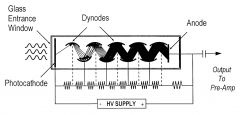
The result of a PMT is that the total “electron multiplication” or gain achieved by the PMT is equal to 5N where N is the number of dynodes. Typically, the PMT gain is on the order of ____.
|
1-10 million
|
|
|
In scintillation detectors (and most other pulse mode detection systems), the signal output by the detector must be ____ before being transferred to the rest of the electronics for amplification and counting.
|
processed
|
|
|
There are two common electronic processing pathways for scintillation counting systems depending on whether the system will be used for ____ or used as a ____ to measure the distribution of event energies.
|
simple counting of events, spectrometer
|
|
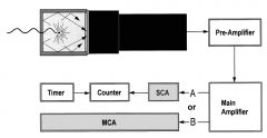
What do "A" and "B" denote?
|
Scintillation detector electronic stage example showing single-channel-analyzer (A) vs. multichannel analyzer (B) electronic pathways.
|
|
|
The first component in the electronic processing pathway of scintillation is the ____.
|
pre-amplifier
|
|
|
The function of the ____ in scintigraphy is to amplify the signal and convert it to a voltage pulse, match the electrical impedance of the detector to the electronic stage to preserve signal, and shape the signal for optimal processing by the electronics
|
pre-amplifier
|
|
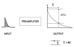
What does this diagram denote?
|
In this example, the pre-amplifier output signal has an exponential decay determined by a simple resistor-capacitor circuit. The time required for the signal to decay to 63% of its maximum is referred to as the time constant τ, and is given by the product of R (resistance in ohms ) x C (capacitance in farads).
|
|
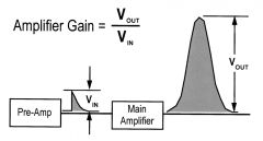
What does this diagram denote?
|
The signal output from the preamplifier has a relatively small amplitude which requires further amplification and shaping and this is achieved by the main amplifier circuit. The amplifier gain is given by the ratio of the voltage amplitude of output signal to that on the input. Amplifier gain is usually adjustable and is on the order of 1-1000.
|
|
Regarding single-channel analyzers, what does this graph represent?
|
In simple counting systems the output from the amplifier is analyzed by a single-channel analyzer (SCA) circuit to determine if the signal amplitude falls within preset upper and lower threshold conditions that correspond to a range of acceptable energies (usually corresponding to the photopeak energy ± some percentage).
|
|
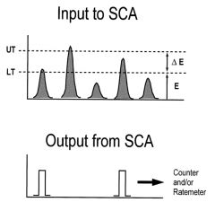
Regarding single-channel analyzers, what does this diagram represent?
|
If the energy of each incoming event falls within the preset limits it is counted until such time that the timer indicates the counting interval is over.
|
|
|
In detectors operated as spectrometers the amplifier output is sent to a multi-channel analyzer which sorts events by energy to form a ____ of energies or energy spectrum, which can be displayed to permit interactive selection of the energy acceptance window to be applied during counting.
|
histogram
|
|
|
Scintillation counting systems can also be operated in a ____ mode where two (or more) detectors are interfaced to a ____ which collects timing information for each radiation interaction in each detector and rejects events that do not happen within a short time interval (____).
|
coincidence, coincidence circuit, coincidence window
|
|
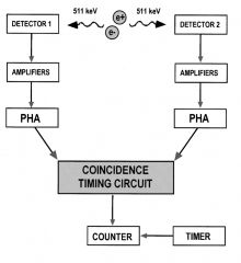
What does the diagram represent?
|
Events occurring within this window are considered to have occurred simultaneously. These detector configurations are often used in counting systems designed for use with positron-emitting radionuclide sources, which after positron emission and annihilation, yield two simultaneous 511 keV annihilation photons traveling in approximately opposite directions.
|
|
|
When radiation interacts in certain scintillation crystals that are phosphors, energy deposited causes electrons to become excited and to move from the ____ to the ____ from where they become trapped in an excited state until some external treatment is applied.
|
valence band, conduction band; There are two common types of scintillators with these properties used in personnel dosimetry in nuclear medicine, the thermoluminescent dosimeter or TLD and the optically-stimulated (OSL) dosimeter.
|
|
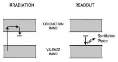
What does this diagram represent?
|
The thermoluminescence process.
|
|
|
____ are small chips or wafers of scintillation crystal that trap and store energy deposited during exposure to radiation and only release a fraction of that energy in the form of scintillation photons after the application of heat when read using a TLD reader.
|
Thermoluminescent dosimeters or TLDs; The readout efficiency for TLDs is typically only a fraction of 1 percent. Before use, TLDs must be heated in an annealing oven in order to release any trapped electrons so they are properly “erased” prior to use.
|
|
|
A common TLD material is the ____ which is used in general dosimeters worn at the collar and ring badges designed for hand dose estimates.
|
LiF chip; TLDs are also used for dosimetry applications using patient-like simulators or phantoms specially designed to accommodate TLD placement in various locations.
|
|
|
Another type of delayed-scintillation detector is the optically-stimulated dosimeter which uses an ____ crystal which traps energy in much the same way that TLDs do.
|
Al2O3; The chips are read by exposing them to light with a wavelength in the green region of the visible spectrum. The subsequent light output is in proportion to the energy deposited in the crystal.
|
|
|
TLDs and OSL dosimeters have many similar characteristics in terms of their application and useful energy range but the ____ dosimeter has the following advantages: it can be repeatedly read (allowing incremental or repeat readings), the readout cycle is faster, and the crystal does not require annealing prior to use.
|
OSL; OSL-based personnel dosimeters have largely replaced film-badges which had been historically used as general purpose personnel dosimeters. As with conventional film badges, the relative absorption through various filter materials provides additional information about the energy and type of radiation to which the dosimeter has been exposed.
|
|
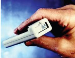
What's this?
|
A solid state dosimeter. The semiconductor diode pocket dosimeter is an example of a solid state dosimeter from which accumulated dose or dose rate readings can be read in real time from an integrated LCD display. Two examples are shown below. These dosimeters have a variety of advanced features including but not limited to activation of an audible alarm when a specified dose limit has been exceeded.
|
|
|
Which direction should a ring badge be positioned?
|
The ring badge should be worn under a glove to avoid contamination and with the label facing the palm of the hand so that the TLD is in closest proximity to the source of activity being handled.
|
|
|
As required by Federal and/or State Regulations, it is mandatory that ____ radiation contamination surveys be conducted in areas where unsealed radioactive sources have been handled.
|
daily; For a typical nuclear medicine clinical operation, this includes but is not limited to: patient dose injection areas, scanning rooms, and the nuclear medicine hot lab or radiopharmacy.
|
|
|
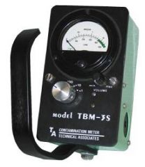
The ____ are more sensitive survey instruments, but only provide an estimate of exposure at the calibration energy of interest. For accurate estimates of exposure, the ____ must be used.
|
G-M detectors, ionization detector
|
|
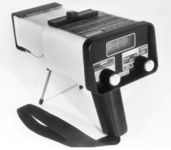
What's this?
|
Ionization detector.
|
|
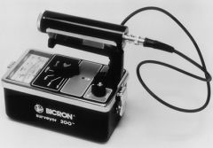
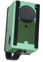
What's this?
|
G-M survey meter
|
|
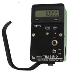
What's this?
|
G-M survey meter
|
|
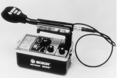
What's this?
|
G-M survey meter with integrated pancake
|
|
What's this?
|
G-M survey meter with integrated pancake
|
|
What's this?
|
G-M survey meter with integrated pancake
|
|
|
The ____ is a high efficiency counting detector used for assessing the relative activity level of in-vitro blood or urine samples collected per established clinical protocols required for hematologic or organ function tests (i.e. blood volume estimates, glomerular filtration rate, etc..).
|
scintillation well counter; When used in this application, small (typically < 0.5 ml) volumes of liquid are placed in narrow plastic counting test tubes and positioned at the bottom of the scintillation detector well.
|
|
|
The well-counter can also be used to monitor ____ acquired when surveying for surface contamination, i.e. on workspace countertops or when checking for leakage of radioactive material (leak test), i.e. on the outer surface of sealed radioisotope sources or on the outer package surface, as well as the inner containment vessel, of incoming radioactive material shipments.
|
“wipe test” samples
|
|
|
When used correctly, the high intrinsic and geometric efficiency of the well counter yields an overall efficiency approaching ____%, nevertheless, well counters are susceptible deadtime losses and therefore should be operated at countrates below ____ counts per second.
|
100, 5,000
|
|
What's this?
|
Well counter detector.
|
|
|
Federal and state regulations require that every dose of radiopharmaceutical to be administered to patients must be measured or assayed prior to administration and this is achieved using a specialized detector called a ____.
|
Dose calibrator.
|
|
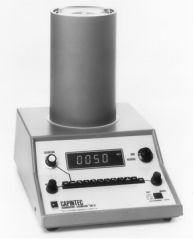
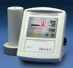
What is this?
|
Dose calibrator.
|
|
|
The thyroid uptake probe is used to estimate the relative uptake of orally-administered I-123 or I-131 by the thyroid gland. It does so by permitting a comparison of the counts obtained in a region overlying the thyroid relative to those obtained with ____.
|
a calibrated capsule of the same radionuclide (I-123 or I-131). The thyroid uptake probe is comprised of a cylindrical scintillation detector coupled to a PMT and is surrounded by a cylindrical collimator to restrict counts to the area of interest. To ensure a relatively consistent probe response, the distance between the patient and probe is kept constant using a positioning reference guide that placed against the patient throat area
|
|
|
More recently, arrays of modular or pixellated CsI(Tl) or other scintillation detectors coupled to silicon photodiodes or position-sensitive photomultiplier tubes have been introduced. These scintillation detectors are comprised of arrays of individual detector elements, each of which is on the order of 3 mm x 3 mm. The use of these pixelated scintillators and the elimination of the conventional PMT array in these new configurations significantly improves ____.
|
spatial resolution.
|
|
|
As required by federal and state regulations, survey meters must be calibrated for accuracy ____ and ____ using a source traceable to a National Institute of Standards and Technology (NIST) standard reference source.
|
annually, after any repair
|
|
|
The accuracy of the survey instrument must be tested at activity levels reflecting points at ____ and ____ of the full activity scale of the instrument.
|
1/3, 2/3
|
|
|
The zero-level of the survey meter must be verified in an area with ____ and then the constancy of the survey meter must be tested with ____.
|
a very low background rate, a long-lived radioactive “check source”
|
|
|
For routine quality control, how often must both the zero-level of the survey meter and constancy be verified?
|
Daily.
|
|
|
The inter-day constancy measurement of a survey meter must be within ± ____%, otherwise the survey meter may need to be recalibrated.
|
5
|
|
|
For those survey meters that are battery-operated, the battery function should also be tested ____.
|
daily
|
|
|
When should the efficiency and energy resolution of the well counter be evaluated?
|
At installation, annually and after repair.
|
|
|
A background reading and constancy test using a dedicated long-lived check source should be performed how often with a well counter?
|
Daily.
|
|
|
For a well counter, the inter-day constancy readings should agree to within ± ____%
|
5
|
|
|
To ensure reliable results, the registration of the well counter pulse height analyzer (PHA) window relative to the photopeak of the radioisotope being assayed should be verified how often?.
|
Prior to each clinical use.
|
|
|
A suspected malfunctioning well-counter (or other counting system) can be evaluated using a ____ which is used to determine if the counting system is behaving as predicted by statistical theory.
|
Chi-Square test
|
|
|
Dose calibrator accuracy testing using one or more NIST- traceable radioisotope calibration standards, is required when?
|
After installation and repair, and on an annual basis thereafter.
|
|
|
In dose calibrators, the measured activity of the calibration standard must be within ± ____% of the decay-corrected certified calibration activity.
|
5
|
|
|
What is a typical source used for dose calibrator accuracy testing?
|
Typically a Cs-137 source (662 keV; T1/2≅ 30 years) or other long-lived source is used for accuracy testing.
|
|
|
Ideally the dose calibrator would also be tested at low and high energies and the radioisotopes ____ and ____ are useful for this purpose.
|
Co-57 (122 keV; T1/2 ≅282 days), Co-60 (1.33 MeV; T1/2≅ 5.3 years)
|
|
|
How frequent is the consistency of performance of a dose calibrator monitored?
|
Daily. It is especially important that once the dose calibrator accuracy has been established, that consistency of performance be monitored on a daily basis. The dose calibrator constancy test is performed using a long-lived source such as Cs-137.
|
|

|
Example of constancy of Cs-137 source activity as measured with a dose calibrator over a 30 day period is shown.
|
|
|
The properly operating dose calibrator will have a ____ response with increasing activity.
|
Linear
|
|
|
What are the two methods to evaluate dose calibrator response?
|
1. Decay Method 2. Attenuator Sleeve Method
|
|
|
For determining dose calibrator linear response, the ____ method requires taking a few hundred millicuries of ____ and assaying it at regular intervals over the course of 3-4 days until the source activity is <10 uCi.
|
decay, Tc99m
|
|
|
For determining dose calibrator linear response, the ____ method is used which employs a series of concentric cylindrical attenuators that are placed inside the dose calibrator to mimic the effects of decay so the measurement can be done in ____ session(s).
|
attenuator sleeve, one
|
|

|
To calculate the linearity of response, a semi-log plot of dose calibrator response over time (or with increasing attenuation) is performed and a linear fit across all measurement points is used in order to verify that each measurement point is within ± 5% of the expected response. Dose calibrator linearity should be verified at installation, after repair, and on a quarterly basis otherwise.
|
|

What are these for?
|
Attenuator Sleeve Method for determining dose calibrator linearity
|
|

What is this calibration for?
|
Though the dose calibrator has been specifically designed for maximum efficiency for its intended application, its response can vary depending on the geometric configuration or relative position of the source being assayed. For this reason it is important to verify the response of the dose calibrator to different source geometries and activity volumes that might be encountered clinically, i.e. with vials and syringes that are routinely used. If the dose calibrator response using a particular source configuration exceeds ± 5% of the reference configuration, a correction factor must be applied to all readings obtained using that particular source/activity geometry.
|
|
|
When is a dose calibrator geometry response test required?
|
At initial installation and after repair.
|
|
|
How often should the energy resolution of the thyroid probe be evaluated?
|
Annually.
|
|
|
How often should the thyroid probe efficiency be tested?
|
At regular intervals depending on the frequency of use and the manufacturer’s recommendations.
|
|
|
A verification of the pulse height analyzer (PHA) window setting and a constancy test using a long-lived check-source should be performed how often on a thyroid uptake probe?
|
Each day the thyroid uptake probe is in clinical use.
|
|
|
How often should an intraoperative probe be tested for constancy?
|
Prior to each use using a dedicated long-lived check source and should be within ± 5% of the expected reading.
|
|
|
For the intraoperative probes with an integrated base unit that allows visualization of the energy spectrum, the pulse height analyzer window should also be verified how frequently?
|
Prior to each use.
|
|
|
An energy integrating detector is operated ____ mode.
|
current
|
|
|
A photon counting detector is operated in ____ mode.
|
pulse
|
|
|
In the scintillation detector the system deadtime is directly dependent on the ____ and ____.
|
design of the electronic stage, the crystal scintillation decay time
|
|
|
What type of interaction? The photon comes in, interacts with the orbital electrons in atom, and kicks one of them out of the atom. The photon’s direction is changed by this event.
|
Compton scatter. The recoil electron has a negative charge so the remaining atom is now ionized and has a positive charge
|
|
|
In Compton scatter, the angle between the direction of the incoming photon and the outgoing photon is known as the ____.
|
scattering angle; The energy of this scattered photon is lower than the incoming photon with the energy difference being transferred to the recoil electron, and that recoil electron may be energetic enough to cause further ionization in the material.
|
|
|
Why type of interaction? A photon comes in and interacts with an orbital electron, but this time all of the photon’s energy is transferred to the recoil electron and the photon completely disappears.
|
Photoelectric effect.
|
|
|
In gamma cameras, how often should the uniformity test and the test with bar phantoms be performed?
|
Uniformity test = Daily before patient scanning
Bar phantom test = Weekly |
|
What does this picture denote?
|
Here are some examples of what happens when gamma camera corrections are not completely or not properly applied. On the lower left, we can see an image that is supposed to be uniform. We can clearly see dark spots where the photo-multiplier tubes are. On the upper left, we can see an image of a bar phantom. The bars in the phantom itself are completely straight, but they don’t appear straight in the image. On the right, however, we can see what these images are supposed to look like when the corrections are properly applied.
|
|
What does this picture denote?
|
If you lose a photo multiplier tube, you can clearly see the result on the uniformity image. One never wants to be in the situation seen on the right, where a photo multiplier-tube has been lost, and the patient image is affected.
|
|
What does the picture denote?
|
Here’s an example of what happens when you do drop things on your gamma camera. You can get a cracked crystal, and that leads to artifacts in the image.
|
|
|
What happens to the field of view and resolution when you move a cone-shaped collimator close to the patient?
|
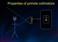
The field of view gets smaller while the resolution increases.
|
|
|
How do you determine the magnification of a collimator?
|

Let’s define the distance from the pinhole to the detector as f, the distance from the pinhole to the patient as b, and then the magnification factor M is given by the ratio of f/b. In this case, f = b, so the magnification factor is 1 and the image is the same size as the object.
|
|
|
How does image distortion occur with collimators and taking advantage of magnification?
|
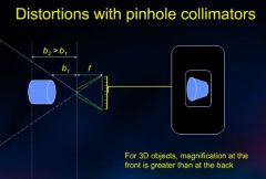
One thing to watch out for is the fact that patients are three-dimensional rather than two-dimensional. The magnification in the front will be greater than that at the back, and that’s going to lead to image distortion. The take-home message is that magnification in small objects is possible with pinhole collimators, but they distort larger objects.
|
|

Regarding sensitivity, what does this diagram demonstrate?
|
A major limitation of pinhole collimators is their poor sensitivity, which is illustrated in this cartoon. Here you can see just how few photons emitted by a point source actually make it through the pinhole to the detector. The sensitivity of a pinhole collimator is strongly dependent on the size of the pinhole. If you increase the pinhole size, then the sensitivity will improve. However, the spatial resolution is going to degrade if you do that.
|
|
|
To summarize, pinhole collimators feature ____ sensitivity, a ____ field of view, and very ____ resolution.
|
low, small, high
|
|
|
With parallel hole collimators, how does sensitivity change with collimator distance from the patient?
|
One might wonder if moving the source closer to the detectors results in a decrease in sensitivity. After all, from further away, more holes will be exposed to the source. However, what we find is that as we get closer fewer holes are exposed, but each one subtends a larger angle. It turns out within a reasonable range the sensitivity using a parallel hole collimator is fairly independent of the distance from the detector. Given that, it really is advantageous to keep the patient as close as possible to the detectors.
|
|
|
What is septal length?
|

Septal length is another property of collimators that has a big impact on spatial resolution and sensitivity. Septal length is defined as the depth of the collimator.
|
|
|
With short septa, what we find is that the spatial resolution is ____ than it would be with the longer septa.
|
poorer; we’re seeing a smaller field of view (per septal space) with longer septa that will result in better spatial resolution.
|
|
|
In parallel collimators, the sensitivity goes down as the septal length is ____.
|
increased
|
|
|
In scintillation cameras, approximately ____% of the absorbed gamma ray energy is converted to light.
|
10
|
|
|
Scintillation cameras typically use how many PMTs?
|
About 55
|
|
|
About how many counts are acquired for a typical scintillation camera image?
|
About 500,000.
|
|
|
What are scintillation camera collimators typically made out of?
|
Lead.
|
|
|
How does the FOV change in parallel-hole collimators as the distance from the patient increases?
|
Does not change.
|
|
|
In converging collimators, how does the FOV change as the camera is moved farther from the patient?
|
FOV decreases with distance
|
|
|
In diverging collimators, they project an image size that is smaller the the object size. How does FOV change with distance?
|
Increases with distance.
|
|
|
High sensitivity collimators have ____ holes and ____ resolution.
|
larger holes and lower resolution.
|
|
|
High resolution collimators have ____ holes and ____ sensitivity.
|
smaller holes and lower sensitivity
|
|
|
What is the most frequent type of collimator used?
|
Low energy, high resolution (LEHR)
|
|
|
Tc-99m and TI-201 use what kind of collimator?
|
LEHR with thin septa
|
|
|
Ga-67 and In-111 use what kind of collimator?
|
Medium energy with thicker septa (and therefore fewer holes and lower sensitivity)
|
|
|
I-131 uses what kind of collimator?
|
High energy (and thickest septa)
|
|
|
Resolution is degraded with ____ distance from the collimator.
|
Increasing
|
|
|
What shape are NaI scintillator crystals, generally?
|
Rectangular
|
|
|
NaI scintillator crystals are usually how thick?
|
10mm
|
|
|
As scinitillator thickness increases, what happens to sensitivity and resolution?
|
Sensitivity improves but resolution decreases.
|
|
|
What is a photopeak?
|
When an incident gamma ray is completely absorbed (photoelectric effect).
|
|
|
What is detection efficiency?
|
The percentage of incident gamma rays abscrobed in the scintillator.
|
|
|
How does increasing the photon energy from 100 to 500 keV affect the detection efficiency?
|
Reduces it from 100% to 6%.
|
|
|
Where does the iodine escape peak occur?
|
Approximately 30 keV below the photopeak.
|
|
|
What causes the iodine escape peak?
|
Is the result of characteristic K-shell xrays from iodine that escape the crystal.
|
|
|
When do scatter events in NaI crystals occur?
|
Scatter events in the NaI crystals occur where the energy of a Compton electron is absorbed in the crystal but the Compton scattered photon escapes.
|
|
|
How much energy is converted to light when the gamma ray is completely absorbed by the NaI?
|
10%
|
|
|
What is the relationship of the output voltage of the photomultiplier tube compared to the amount of energy absorbed by the scintillating material?
|
The output voltage from the photomultiplier tube is directly proportional to the amount of energy absorbed by the scintillating material.
|
|
|
What is photopeak width measured as?
|
Full width half maximum (FWHM)
|
|
|
The broadening of the photopeak (FWHM) is known as what?
|
Energy resolution
|
|
|
What is the electronic device used to determine which portion of the dedicated spectrum is used to create images?
|
Pulse height analyzer
|
|
|
The pulse height analyzer can be set to allow only selected energies to be counted which reduces the number of what type of photons in the image?
|
Compton scatter photons.The pulse height analyzer maximizes the number of photopeak events while minimizing the detected photons that would degrade image quality.
|
|
|
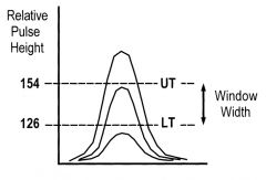
What parameter do you alter in a pulse height analyzer to determine the acceptable range of energies for counting?
|
The window, measured by percent, determines the acceptable range of energies around the peak her subsequent counting. A peak of 140 keV with a 20% window accepts photon energy levels ranging from 126 to 154 keV.
|
|
|
Regarding the pulse height analyzer, what happens to the imaging time when you widen the window?
|
Wide windows accepts more photons and produce images in a shorter time but include more scatter photons that degrade image quality. Some radionuclides such as Gallium-67 require that multiple windows be set since these emit several gamma rays.
|
|
|
What is a typical nuclear medicine matrix size used for cardiac imaging?
|
64 x 64 matrix
|
|
|
Was a typical nuclear medicine matrix size used for whole body imaging?
|
1024 x 1024 matrix.
|
|
|
What is the most commonly used nuclear medicine matrix size?
|
128 x 128 matrix.
|
|
|
Which type of collimator is commonly used for SPECT imaging?
|
Parallel hole collimator.
|
|
|
What is a typical matrix size for cardiac SPECT?
|
64 x 64 matrix.
|
|
|
What is a typical matrix size for noncardiac SPECT imaging?
|
128 x 128 matrix.
|
|
|
What does SPECT currently use, iterated reconstruction or filtered back projection reconstruction algorithm?
|
Scan projections were originally used as inputs or filtered back projection reconstruction algorithms to compute tomographic images. Iterated reconstruction algorithms are now used. Iterated reconstruction is more accurate and minimizes artifact.
|
|
|
What is the major benefit of SPECT imaging compared to planar imaging?
|
Improved contrast that results from the elimination of overlapping structures.
|
|
|
In modern day PET scanners, what are the detectors made out of?
|
lutetium oxyorthosilicate (LSO), gadolinium oxyorthosilicate (GSO), luteitium yttrium oxyorthosilicate (LYSO). GSO is the poorest absorber of 511 keV gamma rays.
|
|
|
Because GSO and LSO have increased light output relative to BSO, what is improved relative to BSO?
|
Improved energy resolution.
|
|
|
Which detector is thicker, planar detectors or PET scan detectors?
|
To efficiently detect 511 keV annihilation photons, fixed detectors are used such as 20 to 30 mm.
|