Reading...
![]()
Play button
![]()
Play button
![]()
Use LEFT and RIGHT arrow keys to navigate between flashcards;
Use UP and DOWN arrow keys to flip the card;
H to show hint;
A reads text to speech;
80 Cards in this Set
- Front
- Back

|
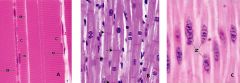
A: Skeletal Muscle – striated, long parallel arrangement, multiple peripheral nuclei
B: Cardiac Muscle – striated, branched cells, central nucleus, Intercalated disks C: Smooth Muscle – fusiform, central “cork screw nucleus” |
|
|
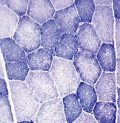
Non-striated muscle type
|
smooth
|
|
|
Skeletal muscle cell (muscle fiber)
Is multinucleated, and formed from fusion of individual ___ during development |
myoblasts
|
|
|
Skeletal muscle cells are long, cylindrical cells (a muscle cell is called a fiber) having ___ nuclei. Each muscle cell is surrounded by a ___
|
-multiple, peripherally located
-basal lamina |
|
|
Nomenclature: (Sarco = flesh)
-Sarcolemma: Sarcoplasm: Sarcoplasmic reticulum: |
-the muscle fiber (cell) plasmalemma
-the muscle fiber cytoplasm -specialized muscle endoplasmic reticulum |
|
|
Endomysium – CT surrounding individual muscle cells, composed of ___ and external lamina (___)
|
-reticular fibers
-basal lamina |
|
|
Perimysium – thicker CT that surrounds and defines ___ (functional bundles of muscle fibers)
|
fascicles
|
|
|
Epimysium – dense CT that surrounds ___ (a collection of fascicles)
|
the entire muscle
Investing fascia of gross anatomy |
|
|
Types of muscle contraction:
___ – not voluntarily controlled ___ – slight contraction that gives muscle firmness, assisting stability of joints and posture |
-Reflexive
-Tonic |
|
|
Types of muscle contraction:
Phasic ___ – muscle length does not change ___ – muscle length changes >>___– contracting muscle shortens during movement >>___ – contracting muscle lengthens during movement (lowering an object or squatting down) |
-Isometric
-Isotonic -Concentric -Eccentric |
|
|
Type I (___, ___, ___) – not easily fatigued (___ muscles/endurance)
___ myoglobin content (oxygen binding protein) ___ mitochondria and ___ in oxidative enzymes ___ myosin ATPase reaction velocity Location: Predominate in the ___ muscles and other ___ muscles of humans to maintain upright posture, soleus muscle of the calf |
Type I (red, slow twitch, slow oxidative) – not easily fatigued (postural muscles/endurance)
High myoglobin content (oxygen binding protein) Many mitochondria and high in oxidative enzymes Slow myosin ATPase reaction velocity Location: Predominate in the deep back muscles and other postural muscles of humans to maintain upright posture, soleus muscle of the calf |
|
|
Type IIb (___, ___, ___) – power and rapid contraction but easily fatigued
___ myoglobin content ___ mitochondria and ___ in oxidative enzymes ___ myosin ATPase reaction velocity Location: ___ |
Type IIb (White, fast twitch, fast glycolytic) – power and rapid contraction but easily fatigued
Low myoglobin content Few mitochondria and low in oxidative enzymes Very fast myosin ATPase reaction velocity Location: extraoccular eye muscles |
|
|
Type IIa ___, ___) they make up ___ motor units that have intermediate characteristics from those described above
|
Type IIa (Intermediate, fast oxidative glycolytic) they make up fast twitch fatigue resistant motor units that have intermediate characteristics from those described above
|
|
|
All muscles are combinations of
|
the 3 fiber types
|
|
|
Does H & E staining reveal fiber type on the basis of color?
|
No; NADH-TR does
|
|
In NADH-TR:
The ___ stain darker. They are ___ in mitochondrial enzymes. They correspond to the red fibers seen in vivo (___). The larger lighter staining fibers correspond with ___ fibers (___ ). |

-smaller fibers
-higher -Type I -white -Type IIb |
|
|
___ are composed of collections of fibers, bound by a perimysium
___ contain collections of myofibrils, which extend the entire length of the cell ___ are composed of myofilaments Myofilaments (2 types): |
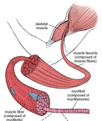
-Fascicles
-Fibers -Myofibrils -Thick filaments & Thin filaments |
|
|
Thin filament:
___ is a double helix polymer of globular monomers of G-actin |

F-actin
|
|
|
___ is a long thin molecule that wraps around and binds to the groove of actin filaments
|

Tropomyosin
|
|
|
Troponin
TnT – TnC – TnI – |
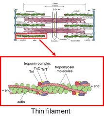
Troponin
TnT – binds to tropomyosin TnC – binds Ca+ TnI – Inhibits actin-myosin binding |
|
|
Thick filaments contain about 250 ___ molecules
|
myosin
|
|
|
Myosin- Composed of 2 identical heavy chains, each
containing a globular head containing binding sites for: ___ also 2 pairs of light chains |
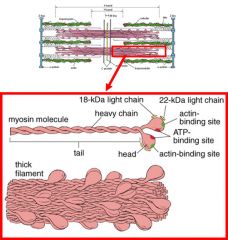
Myosin- Composed of:
-2 pairs of light chains -2 identical heavy chains Contain a globular head containing binding sites for: Actin & ATP |
|
|
|
|
|
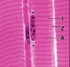
With H&E staining, the A band stains ___ and the I band stains ___ . The Z line is a dark line that ___
|
-darkly
-lightly -bisects the I band |
|
|
A sarcomere is the region between two successive ___. The sarcomere is the ___
|
-Z lines
-functional unit of striated muscle |
|
|
Sliding filament model:
___ filaments slide past ___ filaments ___ bands shorten Sarcomere shortens (Z disks are drawn closer) ___ does not change in length |
-Thin
-thick -I bands and H -A band |
|
|
EM shows that the I band contains ___ filaments only
|
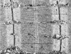
thin
|
|
|
___ contains thick and thin filaments that overlap
|

A band
|
|
|
___ contains only thick filaments, is a light band that bisects the A band and has a darker M line in its center
|

H band
|
|
|
___ is where thin filaments are bound together by the protein ___
|

-Z line
-alpha-actininin |
|
|
___ is narrow, dark region at the center of H bands.
where adjacent thick filaments are linked |
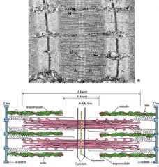
M line
|
|
|
|
|
|
Skeletal Muscle Contraction:
When Ca+ concentration is ___ the binding site on actin, for myosin binding, is blocked |
low
|
|
|
Skeletal Muscle Contraction:
When Ca+ is high, Ca+ binds to ___ , which results in a conformational change that breaks the ___ bond, tropomyosin moves slightly and reveals the binding site on actin for myosin binding |
-troponin C
-troponin I–actin |
|
|
Skeletal Muscle Contraction:
When Ca+ is pumped back into the SR, the cytosolic Ca+ concentration is low again, Tropomyosin returns to its resting state and ___ binding is blocked |
actin-myosin
|
|
|
Skeletal Muscle Contraction Cycle
1. Attachment (rigor) – |
myosin is bound to actin
|
|
|
Skeletal Muscle Contraction Cycle
2. Release - |
myosin is uncoupled from actin and binds ATP
|
|
|
Skeletal Muscle Contraction Cycle
3. Bending: ___ causes myosin head to bend and advance a short distance relative to the thin filament |
Hydrolysis of ATP
|
|
|
Skeletal Muscle Contraction Cycle
4. Force Generation: |
myosin binds to actin and returns to its original conformation – forcing movement of the thin filament along the thick filament (ADP is lost)
|
|
|
Skeletal Muscle Contraction Cycle
5. ___ - cycle can repeat |
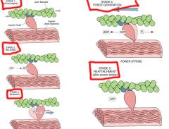
Reattachment
|
|
|
Children with Muscular Dystrophy appear to have large muscular ___, yet they continue to weaken
focal ___ replacement of muscle fibers is seen in the biopsy. This fatty replacement, together with hypertrophic myofibers and increased endomysial fibrosis, is responsible for the pseudohypertrophy seen in the calves of patients with ___. |
-calves
-fat -Duchenne muscular dystrophy |
|
|
Dystrophin – is a cytoskeletal protein located beneath the sarcolema and plays a role in ___ via dystroglycan proteins
|
linking to laminin (ECM protein)
|
|
|
Absense of dystrophin causes ___ (___ disorder) causing progressive muscle weakness in boys
|
-Duchenne’s muscular dystrophy
-X-linked |
|
|
Biopsy from patient, showing the myopathic features:
-___ myofibers -___endomysial fibrosis. |
-atrophic & hypertrophic
-increased |
|
|
Satellite cells between plasma membrane of the muscle fiber and its external lamina serve as ___ for muscle regeneration – this process has limitations
fibrosis occurs to repair injury if the external lamina is disrupted |
stem cells
|
|
|
Triad = SR-TT-SR at the ___ junction
[Note: TT = transverse tubules (T-tubules)] |
A-I
|
|
|
___ act to rapidly spread depolarization throughout the muscle fiber so that there is widespread release of Ca2+ from the SR allowing for uniform contraction of the muscle fiber.
Voltage gated Ca2+ channels open in the sacs of the sarcoplasmic reticulum –releasing Ca2+ into the ___. |
-T-tubules
-sarcoplasm |
|
|
Myelinated axons of motor neurons approach muscle, lose their myelin sheath, and form dilated endings on muscle cells called ___ (3 names)
|
Myelinated axons of motor neurons approach muscle, lose their myelin sheath, and form dilated endings on muscle cells called motor endplates, myoneural junctions or neuromuscular junctions
|
|
|
Axon terminals contain synaptic vesicles filled with the neurotransmitter ___
|
acetylcholine (ACh)
|
|
|
___ in the muscle contain ACh receptors that bind the ACh that crosses the synaptic cleft
|
Junctional folds
|
|
|
___ - a single motor axon and all the muscle fibers it innervates.
Motor units can be very ___ for muscles that require course movements, such as the quadiceps femoris muscle. Or they can be ___, but many in number, in muscles that require fine control such as the extraocular muscle |
-Motor unit
-large -small |
|
|
Myesthenia Gravis
Is an autoimmune disease in which ___. Myasthenia gravis is characterized by |
-antibodies are made to the ACh receptor
-progressive weakness that remits with rest and is worsened by exercise |
|
|
Myesthenia Gravis
___ eyelids and ___ of all muscles of eye movement – no focal deficit |
-Drooping
-weakness |
|
|
Atrophy - shrinkage of muscle fibers
___ can occur in comatose or immobilized patients ___ - muscle cells are dependent on trophic substances supplied by motor neurons and will atrophy following nerve injury |
-Disuse atrophy
-Denervation |
|
|
most common motor neuron disease
|
ALS
|
|
|
ALS:
clinical onset in ___ within several years often leads to death due to ___ ___ atrophy |
-early middle age
-respiratory failure -Denervation |
|
|
Rapid tap of the tendon causes a rapid stretch of the muscle spindle, which initiates the ___ reflex
|
monosynaptic deep tendon
|
|
|
-Muscle spindle is a ___ receptor
-spindle cells (2 types): ___ -(Has afferent and efferent innervation) |
-stretch
-Nuclear chain fiber & Nuclear bag fibers |
|
|
The muscle fibers that produce force for movement of bones termed
|
extrafusal fibers
|
|
|
The spindle cells are termed ___ fibers. They contract to shorten the spindle and retain the ___ of the stretch sensitive region.
|
-intrafusal muscle
-sensitivity |
|
|
CARDIAC MUSCLE:
___ positioned nuclei The fibers (cells) are ___ Intercalated disks form end to end attachments between cardiac ___ |
-Centrally
-branched -myocytes |
|
|
CARDIAC MUSCLE:
T tubules are ___ than in skeletal muscle and are located at a ___ (Not at A-I junction as in skeletal muscle) ___ (SR and T tubule) Large ___ span distance between T tubules |
-larger
-Z disk -Diad -Mitochondria |
|
|
Intercalated disc
Macula adherens (MA – inset #1) Reinforces fascia adherens in the ___ & ___ domain of the disc |
lateral & transverse
|
|
|
Intercalated disc
Gap junctions (GJ – inset # 2) Occupy ___ region of disk |
lateral
|
|
|
Intercalated disc
Fascia adherens (FA – inset #3) Found in the ___ domain and is the major component (staining in H & E) holds cells at their ends Actin filaments attach here |
transverse
|
|
|
Cardiac cells have ___ contractile activity
___ fibers coordinate contraction |
-spontaneous
-Purkinje |
|
|
Purkinje fibers:
Specialized for ___ Have few myofibrils Form ___ junctions with cardiac muscle cells Do not have ___ disks Large pale cells |
-conduction
-gap -intercalated - |
|
|
Myocardial Infarction (MI):
-Death (necrosis) of cardiac muscle cells due to prolonged ___ -Repaired by ___ so function is lost in this region of the heart -___ “cardiac enzymes” is measured to assess damage |
-ischemia (lack of blood)
-fibrous connective tissue -Serum cardiac specific troponin |
|
|
Cardiac mus, distinct characteristics:
each muscle fiber has one or occasionally two, ___ positioned nuclei the fibers (cells) are ___ fibers joined by ___ - specialized junctional complexes between cells that allow cells to act in synchrony (syncytium) via gap junctions ultrastructurally, cardiac muscle has ___ at the Z line rather than triads at the A-I junction |
-centrally
-branched -intercalated disks -diads |
|
|
40% of cardiac muscle cell volume is occupied by ___ versus about 2% for skeletal muscle
|
mitochondria
|
|
|
Smooth Muscle
Non striated and ___ cells Elongated, ___ cells with a ___ centrally located nucleus |
-fusiform
-spindle shaped -single |
|
|
Smooth Muscle
Typically found in |
walls of hollow organs and blood vessels
|
|
|
Smooth Muscle
-organized into sarcomeres? Filaments are loosely organized in association with ___ , which serve same function as Z-line in striated muscle. |
-no
-dense bodies |
|
|
Smooth Muscle
___ is a cytoskeletal intermediate filament protein in all smooth muscle cells. ___ occurs in vascular smooth muscle cells in addition to desmin |
-Desmin
-Vimentin |
|
|
Smooth Muscle
Have no troponin, what is the Ca2+ binding protein? |
CALMODULIN IS Ca2+ binding protein.
|
|
|
Smooth Muscle
Cytoplasmic densities/___ Are proteins (___), which anchor thin filaments and intermediate filaments (Single arrows) Analogous to ___ in striated muscle |
-dense bodies
-alpha-actinin -Z lines |
|
|
Contraction of Smooth Muscle
An increase in cytosolic Ca+ concentration (via gated channels) stimulates smooth muscle ___ Calmodulin binds Ca+ and undergoes a ___ Calmodulin-Ca+ complex activates ___ (phosphorylates one of the myosin light chains) When the globular head of myosin is phosphorylated it interacts with actin and stimulates myosin ATPase Results in ___ Dephosphorylated myosin disassociates from actin Relaxation of muscle |
-contraction
-conformational change -myosin light chain kinase -muscle contraction |
|
|
vascular smooth muscle NN by
|
sympathetic nerves
|
|
|
___ stimulates smooth muscle contraction in the uterus during labor
|
Oxytocin
|
|
|
Vascular smooth muscle is stimulated by
|
passive stretch
|