Reading...
![]()
Play button
![]()
Play button
![]()
Use LEFT and RIGHT arrow keys to navigate between flashcards;
Use UP and DOWN arrow keys to flip the card;
H to show hint;
A reads text to speech;
84 Cards in this Set
- Front
- Back
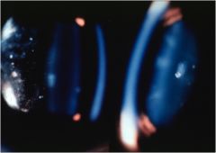
|
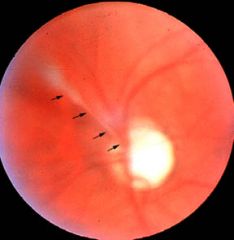
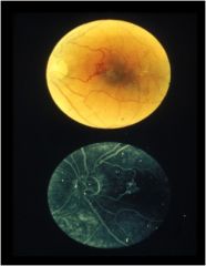
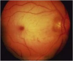
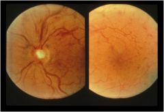
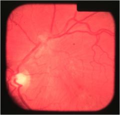
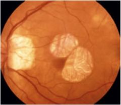
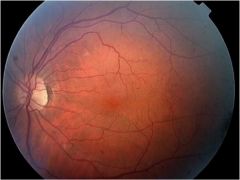
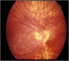
persistent hyloid artery
|
|
|
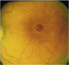
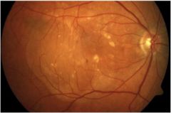
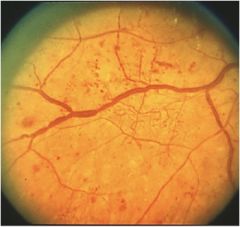
angioid streaks
|
|
|
angioid streaks
|
|
|
asteroid hyalosis
|
|
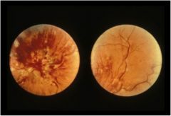
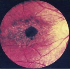
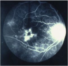
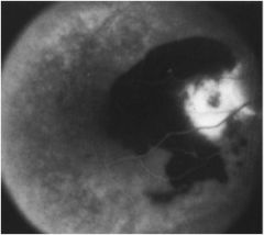
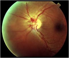
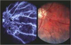
"blood and thunder"
|
CRVO
|
|

|
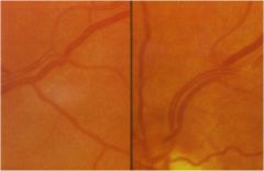
BRAO
|
|
|
BRAO
|
|
|
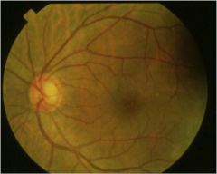
BRVO
|
|
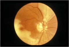
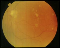
WHAT DO YOU SEE AND WHAT IS THE BLOCKAGE
|
cotton wool, dot blot (i would say flame also) and it is BRVO
|
|
WHAT IS IT AND WHAT DO YOU SEE THAT IS ABNORMAL
|
BRVO with shunt vessels
|
|
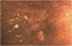
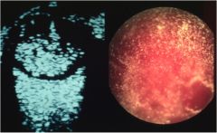
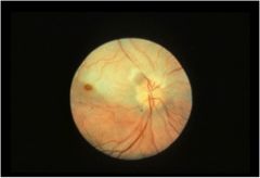
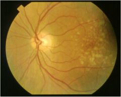
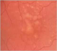
what is it Hint it is not drusen. Only look at the yellow spots, ignore the other defect
|
calcific drusen - notice that it glistens
|
|
|
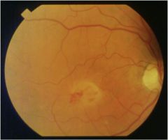
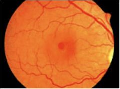
central serous
|
|
|
central serous retinopathy
|
|
what is it and what are the deposits
|
central serous retinopathy with lipid deposits - notice that the deposits are not central
|
|
what is it what is it from
|
choroquine retinopathy from drug toxicities
|
|
obviously their are folds, what layer are they in
|
choroidal folds
|
|
|
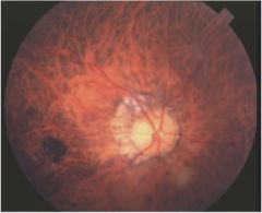
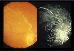
CNV
|
|
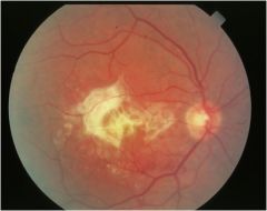
2 things seen here
|
CNV and heme
|
|
notice the CNV, how did it occur
|
CNV from damage that was done to Bruchs membrane from angiod streaks
|
|
drusen or cotton wool
|
cotton wool
|
|
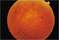
what type
|
CRAO
|
|
WHAT TYPE
|
CRAO
|
|
WHAT TYPE
|
CRAO
|
|
is this an early crao or a brao
|
this is BRAO
|
|
|
CRVO
|
|
THIS PERSON HAD SUDDEN VISION LOSS
|
CRVO
|
|
|
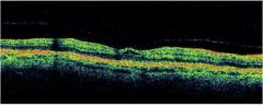
Cystoid macular edema
|
|
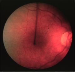
ya its a detachment but what is it from?
|
ROP - retinopathy of prematurity - neo in periphery caused it
|
|
|
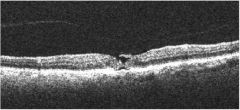
diabetic macular edema
|
|
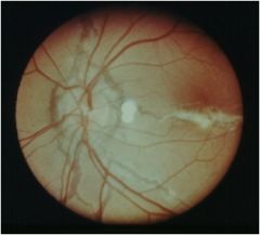
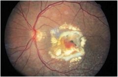
scar named?
|
disciform scar
|
|
scar named?
|
disciform scar
|
|
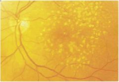
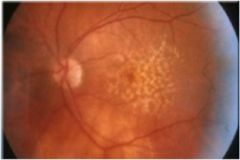
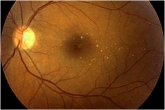
drusen or cotton wool
|
drusen
|
|
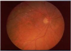
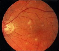
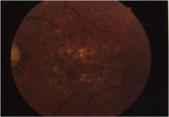
AMD in what phase
|
early
|
|
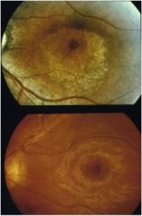
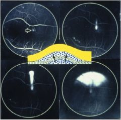
top phase?
bottom Phase? of what? |
top is early bottom late of chloroquine retinopathy
|
|
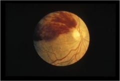
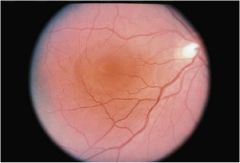
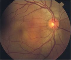
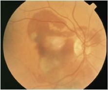
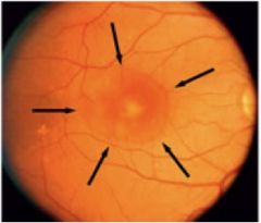
describe what you see
|
elevation (temporal to the macula) with drusen just superior to the elevation
|
|
looks like a nice healthy sheen but this person is 75. what is it
|
epi membrane
|
|
you can see the macular pucker, what is this associated with
|
epi membrane
|
|
this is an epi membrane what is the material over the vessel called
|
glial tissue
|
|
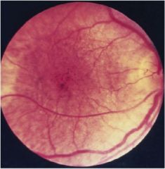
exudative or not?
|
exudative
|
|
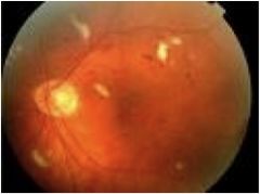
name 2 things seen and what are they from
|
fuchs spot and cresent from myopic degeneration
|
|
|
geographic atrophy
|
|
|
geographic atrophy
|
|
|
geographic atrophy
|
|
|
geographic atrophy
|
|
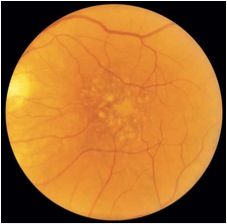
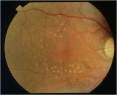
hard or soft drusen
|
hard
|
|
2 problems
|
hypo is heme and the hyper is RPE detachement
|
|
what phase is this hypertensive macular edema in
|
malignant phase with ONH edema
|
|
what AMD phase
|
intermediate
|
|
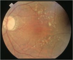
hard or soft druse
|
large area of soft
|
|
what is happening to the vessel
|
lipid sheathing
|
|
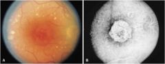
macular hole in what stage?
|
1
|
|
macular hole in what stage?
|
2
|
|
macular hole in what stage?
|
3
|
|
macular hole in what stage?
|
4
|
|
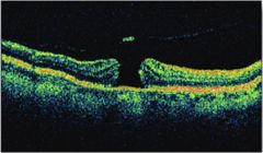
what is it
|
macular hole
|
|
what is it
|
macular hole
|
|
macular pucker from what
|
epi membrane
|
|
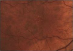
what do you see
|
micro aneurysm
|
|
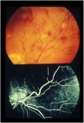
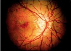
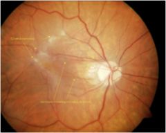
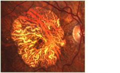
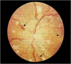
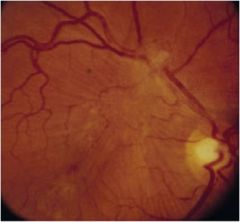
describe what you see
|
neo of the disc
|
|
you can see that there is knicking what would it be called if there was a heme there also
|
Bonnet's sign
|
|
exudative or not
|
exudative
|
|
exudative or non exudative AMD
|
non
|
|
NPDR or PDR
|
NPDR
|
|
NPDR or PDR and what are the white spots
|
NPDR with cotton wool spots
|
|
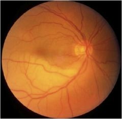
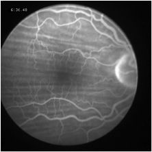
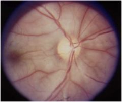
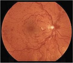
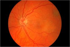
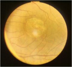
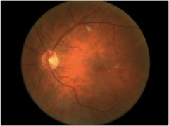
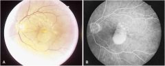
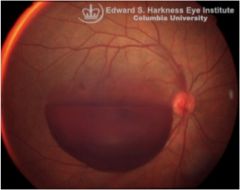
what is it? Hint. notice the location
|
OIS
|
|
|
PED
|
|
|
PED
|
|
what is it what is it caused by
|
PED from confluence of soft drusen
|
|
|
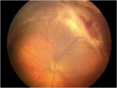
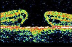
PVD
|
|
HINT: temporal periphery
|
ROP -retinopathy of prematurity
|
|
|
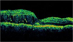
retinoschisis
|
|
|
rubeosis irides
|
|
NPDR in what stage
|
severe
|
|
you know what this is but what is the sign called
|
shaffers sign
|
|
hard or soft or cotton wool
|
soft confluent drusen
|
|
hard or soft
|
soft
|
|
hard or soft
|
soft
|
|
hard or soft
|
soft
|
|
hard or soft
|
soft
|
|
hint its not drusen
|
talc
|
|
hint notice the granularity
|
thioridazine toxicity
|
|
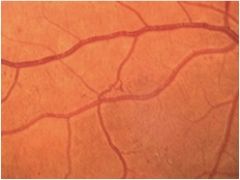
|
venous beading
|
|
NPDR but what do you see
|
venous looping
|
|
what type of heme
|
retro vitreous
|