Reading...
![]()
Play button
![]()
Play button
![]()
Use LEFT and RIGHT arrow keys to navigate between flashcards;
Use UP and DOWN arrow keys to flip the card;
H to show hint;
A reads text to speech;
54 Cards in this Set
- Front
- Back
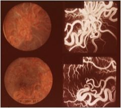
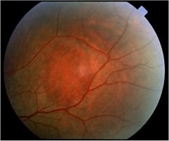
general name?
|
arteriovenous malforations
|
|
|
atypical nevi associated with neurofibromatosis
|
|
|
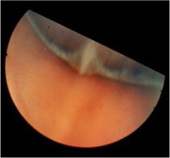
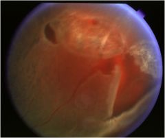
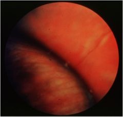
avulsed vitreous base
|
|
|
benign choroidal nevus with drusen
|
|
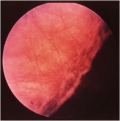
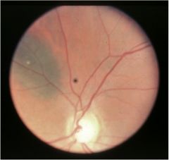
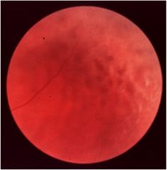
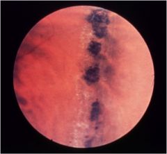
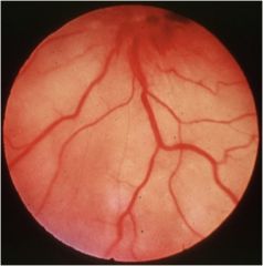
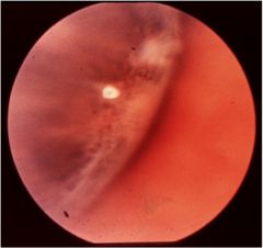
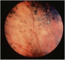
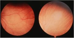
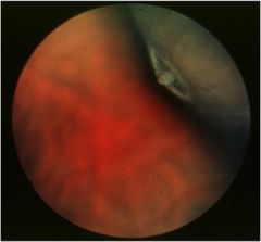
what can you describe about the blood vessels that tells you what area of the eye you are looking at
|
the b/v's begin to turn and go parallel with ora.
|
|
|
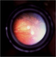
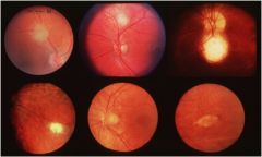
cerebroretinal angiomatosis
|
|
|
cerebroretinal angiomatosis (von hipple lindau)
|
|
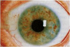
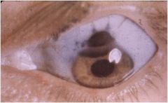
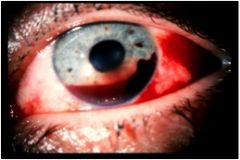
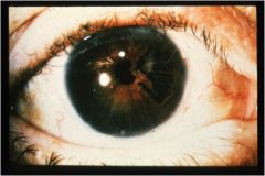
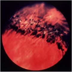
what happened
|
chemical burn - most likely alkali
|
|
|
choroidal nevus
|
|
|
ciliary body malignant melanoma
|
|
|
congenital hypertrophy of retinal pigment epithelium
|
|
|
cystoid degeneration
|
|
hint - looks like honey comb
|
retinoschisis
|
|
DDX from paving stone
|
histo - notice the edges look like an eraser
|
|
name 2 things
|
hole and lattice
|
|
|
horse shoe tear
|
|
|
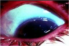
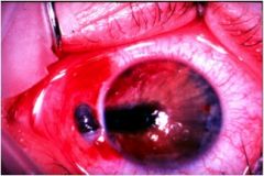
hyphema
|
|
hint it is inferior temporal
|
paving stone
|
|
hint inferior temporal
|
paving stone
|
|
|
irido dialysis
|
|
|
iridoschisis
|
|
|
iris prolaps
|
|
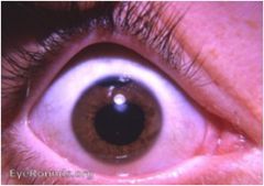
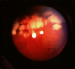
what is this. hint not a disease
|
scars from a laser treatment
|
|
|
lattice
|
|
|
|
lattice
|
|
|
lattice
|
|
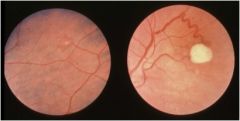
what is it? what are the white lines
|
lattice - lines are blood vessles
|
|

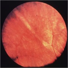
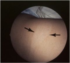
never mind the paving stone. what nerve is that? what is the location?
|
long ciliary nerve on nasal side. you can tell be the blood vessel that is on top running along the superior side of the nerve
|
|
|
melanocytoma
|
|
|
melanocytoma
|
|
|
meridional fold
|
|
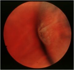
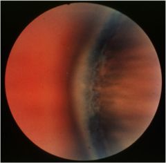
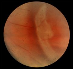
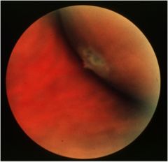
what is it? can you tell if there has been traction?
|
meridional fold yes there is traction you can tell because of the pigmentation
|
|
|
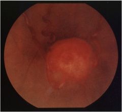
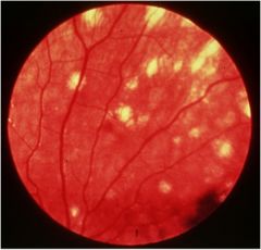
metastatic carcinoma of choroid. it is the creamy white
|
|
|
operculated tear notice the floating disc
|
|
|
oral pearl
|
|
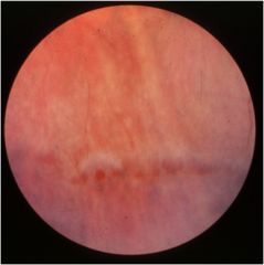
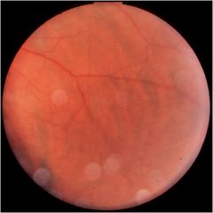
describe location
|
ww/o pressure - parallel to ora
|
|

|
pars plana cyst
|
|
|
paving stone
|
|
|
poor laser tx
|
|
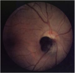
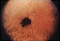
hole or not?
|
pseudohole, this is a pale fundus with a darker area that resembles a hole
|
|
name two things
|
RD and lattice
|
|
|
retinal pigment epithelial hyperplasia
|
|
|
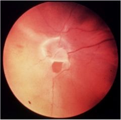
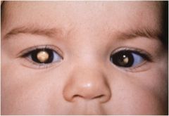
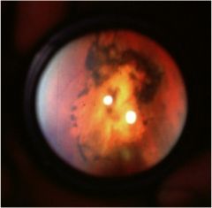
retinal blastoma
|
|
|
retinoblastoma
|
|
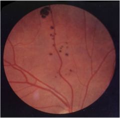
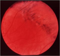
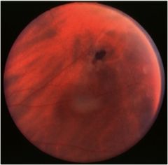
notice blood vessels
|
retinoschisis- the blood vessels in the middle are creating a shadow
|
|
|
short ciliary nerve
|
|
|
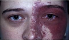
sturge weber syndrome
|
|
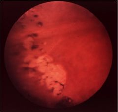
paving stone, histo or toxo
|
toxo
|
|
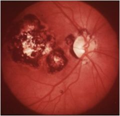
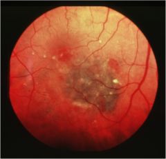
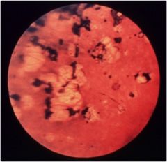
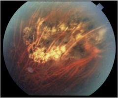
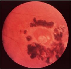
what is it and describe the color
|
toxo with hyperplasia
|
|
hint ash leaf
|
tuberous sclerosis (bournevilles disease)
|
|
|
tuberous sclerosis
|
|
|
vitreoretinal tuft
|
|
|
vitreoretinal tufts
|
|
2 things
|
vitreoretinal tuft surrounded by cystoid degeneration
|