Reading...
![]()
Play button
![]()
Play button
![]()
Use LEFT and RIGHT arrow keys to navigate between flashcards;
Use UP and DOWN arrow keys to flip the card;
H to show hint;
A reads text to speech;
43 Cards in this Set
- Front
- Back
- 3rd side (hint)
|
Axis of the body
Consists of the skull, vertebral column, & rib cage |
Axial Skeleton
|
|
|
|
Supports the appendage or limbs
– Consists of arms, legs, shoulders & pelvic girdle |
Appendicular Skeleton
|
|
|
|
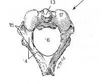
8 cranial bones form the braincase
|
Frontal bone
Parietal bones (2) Temporal bones (2) Occipital bone Sphenoid bone Ethmoid bone |
|
|
|
14 facial bones make up the face
|
Mandible
Maxillary (2) Nasal bones (2) Inferior nasal conchae(2) Lacrimalbone (2) Zygomaticbones (2) Palatine bones (2) Vomer |
|
|
|
Air cavities in the maxillae, frontal, sphenoid, and ethmoidbones
lightens the skull & provides resonance for voice |
Paranasal sinuses
|
|
|
|
Air cavities in the mastoid process of temporal bone
|
Mastoid sinuses
|
|
|
|
Bones of the spine (33)
|
7 Cervical
12 Thoracic 5 Lumbar 5 Sacral 4 Coccygeal |
|
|
|
Number of curves in the spine
|
4 = L,K,LK
|
|
|
|
PRIMARY CURVE LOCATED IN: Thoracic &
Sacrococcygeal convex posteriorly concave anteriorly an alignment |
kyphosis
|
|
|
|
PRIMARY CURVE LOCATED IN:
Cervical & lumbar convex anteriorly Concave posteriorly an alignment |
LORDOSIS
|
|
|
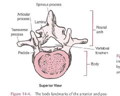
WHAT DOES THE NEURAL ARCH DO?
|
protects the spinal cord and its associated nerve roots
|
|
|

WHAT DOES THE VERTEBRAL FORAMEN DO?
|
allows for the passage of the:
spinal nerve root dorsal root ganglion spinal artery of the segmental artery communicating veins between the internal and external plexuses, recurrent meningeal(sinu-vertebral) nerves, and transforaminalligaments. |
|
|
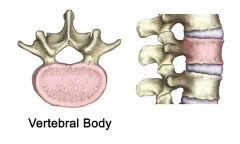
WHAT DOES THE VERTEBRAL BODY DO?
|
bears about 80% of the load while standing and provides an attachment for the discs between the vertebrae.
|
anterior section
protects the spinal cord and nerve roots. |
|
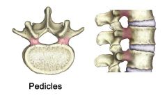
WHAT DO THE PEDICLES DO?
|
providing side protection for the spinal cord and nerves.
serve as a bridge, joining the front and back parts of the vertebra |
|
|
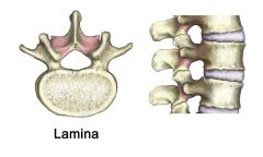
WHAT DOES THE LAMINA DO?
|
roof of the spinal canal that provides support and protection for the backside of the spinal cord
|
|
|
NAME THE STRUCTURE AND WHAT IT DOES.
|
above and below the pedicles are named the vertebral notches
|
when the vertebrae are PUT TOGETHER, the notches of each PAIR ARE CALLED
intervertebralforamina |
|
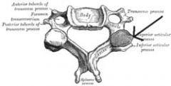
NAME THE STRUCTURE AND ITS FUNCTION
|
•two superior and two inferior, spring from the junctions of the pedicles and laminæ.
•These stick out of an end of a vertebra to lock with a zygapophysison the next vertebra, to make the backbone more stable. |
|
|
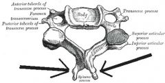
WHAT DOES IT DO?
|
LOCATED junction of pedicles and the lamina
provide a place for the back muscles to attach to the spine absent in the vertebra of the neck (the cervical spine) |
If present in the cervical spine they occur at the lowest level (C7) and are called a cervical rib
may impair exiting nerve roots and cause pain |
|
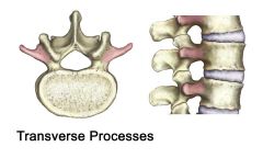
WHAT STRUCTURE IS THIS AND WHAT DOES IT DO?
|
directed backward and downward from the junction of the laminae(in humans), and serves for the attachment of muscles and ligaments
|
|
|
WHAT DOES IT DO?
|
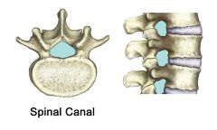
a bony tunnel surrounding the spinal cord.
•It is made up of the front (anterior) of the vertebral body, the pedicles on the sides of the vertebral body and the lamina in the back. •In the lower back it also contains the nerve roots of the lower spine. |
|
|
WHAT DOES IT DO?
|
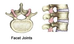
allowing a connection with the vertebrae above and below it
*The pair that faces upward is the superior articularfacet. •The pair that faces downward is the inferior articularfacet. •The facet complex is surrounded by a watertight synovial capsule, much like the small joints in the fingers that allow for smooth movement. |
|
|
NAME THE STRUCTURE AND WHAT IT DOES.
|
comprises a bony tube like vertebral canal that encloses the spinal cord and its meninges.
|
|
|
What is the structure and function?
|
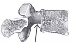
Atlas (1stvertebrae)
– Supports the cranium – Has no body but resembles a bony ring – Transverse process protrudes more laterally; – Spinousprocess is shortened; – Articularfacets: superior are concave; inferior are flat to slightly concave |
|
|
|
Name structure, location, and function.
|
Axis (2ndVertebrae)
– Spinous process is strong & prominent & easy to identify – It is the pivot around which the first vertebra rotates – Odontoid process (Dens) where cervical rotation occurs through its articulation with the atlas |
|
|
Name structure, location, and function.
|
Axis (2ndVertebrae)
– Spinous process is strong & prominent & easy to identify – It is the pivot around which the first vertebra rotates – Odontoid process (Dens) where cervical rotation occurs through its articulation with the atlas |
|
|
|
C3 TO C6
|
Facets are oriented at a 45°angle to the transverse plane
– Guide motion but do not limit it. |
|
|
|
•
Support the chest • Serve for articulation of the ribs • All vertebrae exhibit four articularfacets which form the articulations with the 12 ribs |
THORACIC AREA
|
|
|
|
The facets of the spine are oriented at a ___°angle to the transverse plane and a ___°angle to the frontal plane.
|
60, 20
|
|
|
|
Spinous processes project ______especially the 2nd-10th
• A characteristic which limits the ability of the thoracic spine to hyperextend. |
DOWNWARD
|
|
|
|
•
Carry most of the weight of the trunk • Articularprocesses are more defined & provide more of an interlocking articulation which limits movement around the vertical axis |
LUMBAR
|
|
|
|
IN WHAT PART OF THE SPINE...
The facets are oriented at a 90°angle to the transverse plane and a 45°angle to the frontal plane |
LUMBAR AREA
• |
|
|
|
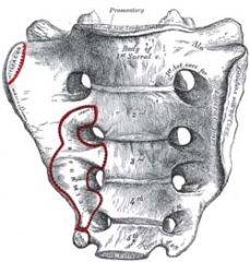
•
Place at which the pelvis is anchored • Represents the fusion of five sacral vertebrae to form a large triangular bone located like a wedge between the two hip bones • No movement is possible in this area |
SACRUM
|
|
|
|
Provides movement between the 1st& 2ndcervical vertebrae to provide a pivoting rotation movement
– Referred to as a “screw joint” – Approx. 50% of the rotation in the cervical spine occurs here |
•
Atlanto-axial (C1-2) – |
|
|
|
WHAT JOINT?
Two degrees of motion – Provide movement between the head & vertebral column – Movement is mainly nodding in the sagittal plane – Approx. 50% of the head flexion-extension that occurs in the cervical spine occurs at this joint |
Atlanto-occipital
|
|
|
|
Are classified as fibrocartilaginous
– Account for approx. 25% of the length of the vertebral column |
Intervertebral disk joints
|
|
|
|
C2-S1 vertebrae are separated & flexibly bound together by intervertebral disks
– Allow small motion in all directions |
•
Intervertebral Joints – |
|
|
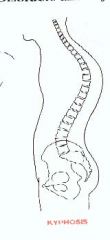
WHAT TYPE OF PATHOLOGICAL CURVATURE?
|
Kyphosis
– Increase in the posterior convexity of the thoracic curve (hump back) – Frequently related to osteoporosis |
|
|
|
Lateral deviation of the spine
– May be functional or structural – More common in females; it becomes apparent in the teen years – Caused by unequal development of the vertebral muscles |
Scoliosis
|
|
|
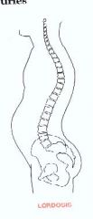
EXPLAIN LORDOSIS
|
Lordosis
– Excessive increase in one of the forward convexities of the normal vertebral column – Lumbar lordosis or cervical lordosis – Frequently referred to as “swayback” – Pregnancy often develops a temporary lumbar lordosis |
|
|
|
MOST FLEXIBLE
|
CERVICAL NECK REGION
|
|
|
|
The vertebral column has___degrees of freedom of motion.
|
6 DEGREES
|
|
|
|
HUNCHBACK
|
KYPHOSIS
|
|
|
|
SWAYBACK
|
LORDOSIS
|
|