![]()
![]()
![]()
Use LEFT and RIGHT arrow keys to navigate between flashcards;
Use UP and DOWN arrow keys to flip the card;
H to show hint;
A reads text to speech;
92 Cards in this Set
- Front
- Back
|
Oral CHO digestion in monogastrics: mechanisms and what they do |
Mastication - decreases particle size and increases area for enzyme digestion Salivary amylase - Converts starch to maltose and smaller oligosaccharides |
|
|
In which species does Amylase has a minor role in CHO digestion? |
Pigs and Dogs |
|
|
Corpus and Fundus CHO Digestion for monogastrics: mechanisms and how they work |
Salivary amylase converts starch to maltose and smaller oligosaccharides (*important in pigs)
(amylase rapidly inactivated by low pH) |
|
|
Intestinal CHO digestion in monogastrics |
Pancreatic amylase converts remaining starch and polysaccharides to monosaccharides |
|
|
What percentage of starch breakdown occurs in the: Oral cavity? Corpus/Fundus? Intestines? |
Oral: ~5%
Corpus/Fundus: ~30%
*Remainder in the intestines |
|
|
Phases of intestinal digestion |
Luminal phase
Membranous phase |
|
|
Luminal Phase: -Where are the enzymes active? -What occurs during this phase (overview)? |
Enzymes active in gut lumen
Large polymers like starch and protein digested by digestive enzymes from salivary, gastric and pancreatic glands |
|
|
Membranous Phase: -Where are the enzymes active? -What occurs during this phase (overview)? |
Enzymes active at surface of gut
Small polymers like polysaccharides and peptides digested by digestive enzymes synthesized in enterocytes and attached to apical membrane |
|
|
Specific starch digestion phases in the intestine |
Pancreatic Phase
Mucosal Phase
Delivery Phase |
|
|
What occurs during pancreatic phase? |
alpha-amylase from pancreas breaks down starch into glucose (first goes through a-dextrine, maltotriose, maltose) Lactase breaks down lactose into galactose and glucose |
|
|
What occurs during mucosal phase? |
Glucose is taken up into intestinal epithelium by the transporter: SGLT1 on brush border
Glucose can also move through paracellularly (paracellular diffusion) |
|
|
What happens in delivery phase? |
Glucose in the epithelial cell undergoes metabolism for cell functions
Glucose gets transported out of cell and into the portal circulation through the transporter: GLUT2 |
|
|
Where does the energy for the SGLT1 come from? |
Na-K ATPase transporter |
|
|
Name sodium-glucose co-transporters |
SGLT1 SGLT2 SGLT3 |
|
|
SGLT1: -Found in which major tissues? -Functions? |
Kidney, Intestine
Glucose reabsorption in intestine and kidney |
|
|
SGLT2: -Found in which major tissues? -Functions? |
Kidney**
Low affinity but high selectivity for glucose |
|
|
SGLT3: -Found in which major tissues? -Functions? |
Small Intestine, Skeletal muscle
Glucose activated Na+ channel |
|
|
Facilitative Glucose Transporters: name the important ones |
GLUT1, GLUT2, GLUT4, GLUT5 |
|
|
GLUT1: -Found in which major tissues? -Functions? |
Ubiquitous: RBCs, Brain, Eye, Mammary
Basal glucose uptake and transport across blood tissue barrier |
|
|
GLUT2: -Found in which major tissues? -Functions? |
Liver, Kidney, Pancreas, Small Intestine
High capacity, low-affinity transport |
|
|
GLUT4: -Found in which major tissues? -Functions? |
Muscle, Fat, Heart
Insulin-regulated transport in muscle and fat |
|
|
GLUT5: -Found in which major tissues? -Functions? |
Intestine, Kidney, Testis
Fructose transport |
|
|
What are CHOs good for in Ruminants |
Major energy source -50% of grain fed to animals -60-70% total diet in dairy cattle |
|
|
Main functions of CHOs in ruminants? |
Energy for microbes and host Maintain a healthy GIT |
|
|
CHO fermentation productions in ruminants? |
VFAs: Acetate, Proprionate, Butyrate |
|
|
Ruminal digesta fractions and what they contain |
1. Fibrous raft - high density of microbes 2. Liquid fraction - shuttles saliva & fermentation end products in and out 3. Boundary layer against lumen surface - bidirectional fluid exchange 4. Soupy Material - provides cud and material for omasum |
|
|
Where is soupy material found? |
Ventral and cranial sacs |
|
|
Define: Fermentation |
A slow, digestive process of anaerobic digestion by microbial enzymes |
|
|
Synergy between host and microbes: what does each provide the other? |
Host provides environment (substrate, H20, pH, redox conditions, slow digesta flow)
Microbes convert indigestible material into useful products for host (VFAs, CH4, NH3, CO2, Microbial cells and LCFA) |
|
|
What are the requirements for fermentation? |
1. Substrate (regular supply of new feed) 2. Microbes (suitable numbers and types) 3. Mixing and propulsion (helps fermentation) 4. Fermentation end products (steadily removed) 5. Stable intraruminal conditions |
|
|
What are stable intraruminal conditions? |
Optimum temperature: 37'C Osmolality: 300 mOsm pH: 6.4 Anaerobic conditions: most microbes are strictly anaerobic; -ve oxidation reduction potential |
|
|
Rumen protozoa: digestion and fermentation |
-Engulf feed particles, digest and store CHO, proteins and fat -Ingest bacteria -Produce some VFAs and NH3 -Slow digestion of rapidly fermentable starches and proteins |
|
|
Rumen protozoa decrease significantly when? |
High grain diets are fed and pH <5.5 |
|
|
Advantages of rumen protozoa |
1. Store starch particles, thereby preventing rapid degradation by amylolytic bacteria 2. Reservoir of microbial protein 3. Provide host with protein, stored starch and PUFA when digested |
|
|
Types of rumen bacteria based on location |
1. Free-living in the liquid phase 2. Loosely/firmly associated with feed particles 3. Associated with rumen epithelium 4. Attached to surface of protozoa and fungi |
|
|
Types of rumen bacteria based on fermentation |
1. Primary bacteria
2. Secondary bacteria |
|
|
Primary bacteria: What do they do? Examples? |
Directly break down digesta -Cellulytic (18h doubling time, slow metabolism, Opt. pH ~6.5) -Amylolytic (<4h doubling time, Opt. pH 5.5-6.8) |
|
|
Secondary bacteria: What do they do? Examples? |
Feed on by-products of primary bacteria (16h doubling time, Optimum pH 6.2-6.8) Propionate bacteria - utilize lactate Mathanogenic bacteria - utilize H2 |
|
|
Process of rumen bacterial attachment to plants |
1. Free bacteria move to fibrous substrate 2. Initial non-specific adhesion on cut surfaces (electrostatic/ionic) 3. Specific adhesion (ligands/adhesins on bacterial surface) *Proliferation and colonization on feed |
|
|
What happens to cellulose digestion if unsaturated fat is fed? |
It leads to decreased cellulose digestion due to interference of bacterial attachment to substrate |
|
|
Benefits of bacterial attachment to plants |
1. Brings enzyme & substrate together 2. Protects enzymes from proteases in rumen 3. Allows bacteria to colonize the digestible surface of feed particles 4. Retention in the rumen to prolong digestion 5. Reduces predatory activity of protozoa |
|
|
Function of protozoa |
Ingest and digest feed particles producing VFA, CO2 and NH3
Also keeps bacteria in check |
|
|
Protozoal storage |
Feed particles like CHO, fat and protein to prolong digestion |
|
|
Location of protozoa |
Both liquid and solid phases |
|
|
Number and size of protozoa |
10^4 - 10^6 cells/gm of rumen contents ~51% of microbial volume |
|
|
pH sensitivity of protozoa |
Highly pH sensitive so they decreases in number on high grain diets |
|
|
Function of bacteria |
Ferment CHO, fat and protein to produce VFA, CO2 and NH3 |
|
|
Bacterial storage |
Very limited/no storage capacity |
|
|
Location of bacteria |
Free living in rumen fluids, or Loosely/firmly attached to feed particles, or Attached to rumen epithelium (mainly fungi for fibre digestion) |
|
|
Number and size of bacteria |
10^10 - 10^12 cells/gram of rumen contents ~40% of microbial volume
*much smaller in size than protozoa |
|
|
pH sensitivity of bacteria |
Some strains are pH resistant |
|
|
In a high forage: low concentrate diet, which VFAs are produced and relative quantity?
In a low forage: high concentrate diet, which VFAs are produced and relative quantity? |
Mainly acetic acid, some acetic acid, the least butyric acid
Mainly propionic acid, some acetic acid, the least butyric acid
*butyric acid stays constant |
|
|
Which VFA is the gluconeogenic one? |
Propionate |
|
|
How are VFAs transported from rumen fluid into the epithelial cells? |
1. Simple diffusion (butyrate) 2. HCO3-/SCFA- antiporter 3. Lactate/H+ symporter |
|
|
What happens in the rumen epithelial cell after VFAs enter? |
Some HSCFA get broken down into SCFAs and then into KETONES |
|
|
Main transporter for ketones from rumen epithelial cell into the blood? |
MCT1 |
|
|
Methods of VFA absorption into the blood (across basolateral membrane)? |
1. Simple diffusion (HSCFAs) 2. Cl-/SCFA- antiporter 3. HCO3-/SCFA- antiporter 4. MCT1 |
|
|
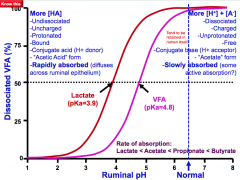
VFA disassociation curve |
|
|
|
4 stages of VFA synthesis |
Hydrolysis Glycolysis VFA synthesis Microbial protein synthesis |
|
|
Precursors for: Acetate Butyrate Propionate |
Phosphenol pyruvate
Acetyl CoA
Oxaloacetate & Lactate |
|
|
Amount of Ruminal epithelial metabolism of VFAs and what it produces |
90% Butyrate --> Ketone bodies (alimentary ketogenesis)
70% Prpionate --> lactate + CO2 + amino acids
30% acetate |
|
|
Energy production, in ATP/mol, for each VFA |
Acetate: 10
Propionate: 18
Butyrate: 27 |
|
|
Which VFAs are not used for net glucose synthesis? |
Acetate and Butyrate |
|
|
What does acetate provide? |
Carbon source for fatty acids in adipose and mammary gland |
|
|
What does propionate provide? |
Glucose Stimulates INSULIN secretion 5% converted to lactate |
|
|
What does butyrate provide? |
Carbon source for fatty acids in mammary gland Stimulates INSULIN and GLUCAGON secretion Critical for rumen papillae growth |
|
|
What is Parakeratosis?
What are events leading up to it? |
Excessive keritinization of the epithelium
Increase in fermentable CHO feeding initially causes sloughing of stratum corneum leading to parakeratosis and formation of stellate ulcers |
|
|
Major type of bacterium in animals fed high grain diets |
Amylolytic |
|
|
Examples of Ruminal bacteria that rapidly ferment starch/soluble sugars |
Selenomas ruminantium Streptococcus bovis Lactobacillus species |
|
|
Describe S. ruminantium |
Dominant organism that makes up 22-51% of culturable bacteria in the rumens of high-grain diet animals |
|
|
Describe S. bovis |
A facultative anaerobe that has explosive growth in response to availability of fermentable CHO especially when animal unadapted to high grain Has a rapid growth rate and rapidly degrades cereal grain starches |
|
|
Lactic acid production by S. bovis causes what? |
Decrease in ruminal pH which inhibits growth rates of most ruminal bacteria This causes acid-tolerant Lactobacilli to predominate |
|
|
Describe lactobacilli |
Predominant organism in acidotic rumens Lactic acid producing! |
|
|
Sequellae of rumen acidosis |
Rumenitis (stellate ulcers) Liver abscesses (F. Necrophorum) Diarrhea due to increased osmolality of fluids Laminitis Sudden death from dehydration, acidosis, endotoxemia |
|
|
Glycogenesis - Glycogenolysis: Liver vs. Muscle |
Liver has glucose-6-Pase enzyme to produce glucose Muscles don't have enzymes to sythesize glucose from lactate so glycolysis occurs and lactate then transported into liver *Liver takes up lactate and undergoes gluconeogenesis to produce glucose |
|
|
Rate limiting enzymes for glycogenesis |
Glycogen synthetase |
|
|
Rate limiting enzymes for glycogenolysis |
Glycogen phosphorylase Glucose-6-Phosphatase (G-6-Pase_ |
|
|
What happens during Cori cycle? |
Lactate from muscle is transported into liver via blood - Gluconeogenesis from lactate in liver - glucose returned to muscle via blood |
|
|
Sources for gluconeogenesis for ruminants |
Propionate Amino acids Lactate Glycerol |
|
|
In ruminants, gluconeogenesis meets ________ of total glucose needs |
75-90% |
|
|
Gluconeogenesis sources in carnivores |
Amino acids |
|
|
Gluconeogenesis sources during anaerobic exercise |
Muscle lactate (Cori cycle) |
|
|
Plasma glucose differences between ruminants and carnivores |
50-60mg/dl in ruminants (very stable levels)
100mg/dl in carnivores (fluctuations after meals) |
|
|
Principle types of Hypoglycemia |
1. Insufficient gluconeogenesis & glycogen stores 2. Mesenchymal tumors 3. Glycogen storage diseases 4. Hyperinsulinemia |
|
|
Insufficient gluconeogenesis and glycogen stores most common in? why? |
Ruminants during pregnancy and lactation Neonatal pigs
Adrenal cortex/anterior pituitary deficiency Liver diseases |
|
|
Why do Mesenchymal tumours cause hypoglycemia? |
Inhibits glucagon |
|
|
Hyperinsulinemia: how does it occur (pathway)? Name phenomenon? |
Somogyi overswing Insulin overdose - hypoglycemia - increase glucagon and cortisol secretion - hyperglycemia and glucosuria |
|
|
Hyperinsulinemia in dogs |
Organic: Islet tumours
Functional: Insulin secretion normal at fasting, but hypersecretion after CHO ingestion |
|
|
Principle types of hyperglycemia |
Diabetes Mellitus -Type 1: juvenile or IDDM -Type 2: adult onset or NIDDM -Other types |
|
|
What causes Type 1 DM |
Primary insulin deficiency Beta-cell failure (autoimmune disorder) Common in dogs and cats |
|
|
What causes Type 2 DM |
Primary insulin resistance Beta-cell exhaustion and atrophy Typical in humans, less common in dogs and cats Early lactation cows can have it too |
|
|
Other types of DM |
Gestational diabetes Diestrus diabetes |