Reading...
![]()
Play button
![]()
Play button
![]()
Use LEFT and RIGHT arrow keys to navigate between flashcards;
Use UP and DOWN arrow keys to flip the card;
H to show hint;
A reads text to speech;
38 Cards in this Set
- Front
- Back
|
Why is the SCM called such?
|
Bc it has attachments at the sternum, clavicle, and mastoid process.
|
|
|
What nerve innervates the SCM? What other muscle close by is innervated by this nerve? Is sensory information/pain sent through these nerves?
|
CNXI or Accessory nerve. Also innervates the trapezius. Sensory afferents do not travel through the accessory nerve (CNXI), but rather through C2, C3 nerves.
|
|
|
If the trapezius and SCM flex, how does the head move? What if you have accessory damage on the right side?
|
There's a lateral flexion to the same side and rotation to the opposite side. R side damage to accessory n causes trouble turning the head to the left. In other words, the head turns to the side of the lesion. (Try it turning your head to the left with resistance - the right SCM is flexed)
|
|

What nerve is poking through between the anterior and intermediate scalene? Describe the insertion and attachment points for the scalenes.
|
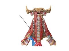
Phrenic N. Anterior, intermediate, and posterior scalenes attach to the transverse processes of the cervical vertebrae. The anterior and intermediate scalenes attach to rib 1. The posterior scalene attach to rib 2.
|
|
|
What are the four Suprahyoid Muscles and what are they innervated by? (N68)
|
1. Myohyoid (CNV3 - mandi div)
2. Digastric (anterior innervated by CNV3 and posterior innervated by CNVII) 3. Stylohyoid (CN VII) 4. Geniohyoid (C1-2) (N68) |
|
|
What are the four Infrahyoid Muscles and what are they innervated by?
|
1. Omohyoid
2. Sternohyoid 3. Sternothyroid 1-3 innervated by the ansa cervicalis 4. Thyrohyoid (C1 via hypoglossal n; CN XII) |
|
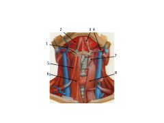
Name these structures. What is not shown below 2?
|

1. Stylohyoid
2. Mylohyoid 3. Digastric 4. Digastric 5. Omohyoid 6. Sternohyoid 7. Thyrohyoid 8. Sternothyroid Below the Mylohyoid is the Geniohyoid |
|

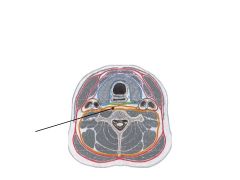
Arrow point to ?? AKA ?? What muscles does it contain?
|
Superficial AKA investing fascia (Called such because it invests the SCM and trapezius mm).
|
|

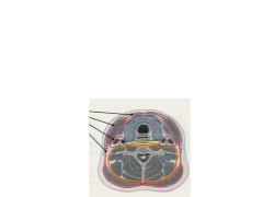
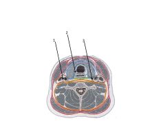
What are the fascia layers called?
|

Red: Superficial/Investing layer
Purple: Muscular Blue: Visceral layer Green: Buccopharyngeal Brown: Carotid sheath Orange: Prevertebral |
|

What does the muscular layer invest?
|
The supra- and infra-hyoid muscles.
|
|

What structures does the visceral layer invest? What is it continuous with?
|
Thyroid gland, trachea & esophagus. Continuous with the buccopharyngeal fascia
|
|

What does the buccopharyngeal fascia separate?
|

It separates the esophagus from the prevertebral fascia.
|
|

What fascial layers make up the "pretracheal fascia"?
|

The muscular, visceral and buccopharyngeal fascia.
|
|
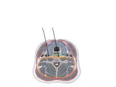
What fascia is the arrow pointing to? What does it attach to?
|

The alar fascia. It attaches to the anterior tubercles of the transverse processes.
|
|

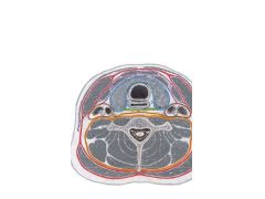
Name the red, blue, green, orange and orange/red layers.
|
Red = investing
Blue = visceral (pretracheal) Green = bucchopharyngeal Orange = alar Red/Orange = prevertebral |
|
|
Where does the alar fascia become continuous with the pre-tracheal fascia?
|
Between T1 and T2
|
|

The carotid sheath is really made up of three fascia = what are they? What three things does it contain?
|

1. Anteriorly it includes the investing and pretracheal layers. Posteriorly, it includes the prevertebral layer of deep cervical fascia.
2. Investing structures include the common and internal carotid arteries, the IJV, and vagus n (CNX). |
|

Name the nerves
|
1. Vagus
2. Recurrent laryngeal 3. Sympathetic trunk |
|
|
What is "I see 10cc in the IV"?
|
How to remember what's in the carotid sheath:
I see = Internal Carotid 10 = vagus n CC = common carotid IV = Internal Jugular Vein |
|
Name these spaces
|

1. Retropharyngeal space
2. Prevertebral space (danger space #4) |
|
|
Describe how the prevertebral space runs compared to the retropharyngeal space.
|
Prevertebral space travels all the way down to the diaphragm. Retropharyngeal travels down only to the T1/2 space - not the thorax - unless it eats through that space and into the prevertebral space.
|
|
|
What are the posterior and anterior triangles of the neck? Can you draw your diagram?
|
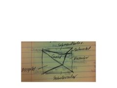
Posterior:
1. Occipital 2. Subclavicular Anterior: 1. Submandibular 2. Submental 3. Carotid 4. Muscular |
|
|
What borders the occipital triangle?
|
Trapezius, sternocleidomastoid, inferior belly of omohyoid.
|
|
|
What are the contents of the occipital triangle?
|
Jugular, cervical plexus, accessory n (CN XI), trunks of brachial plexus, transverse cervical artery, cervical lymph nodes
|
|
|
What borders the omoclavicular triangle (subclavian)?
|
Inferior boarder of omohyoid, clavicle bone, SCM.
|
|
|
What are in the contents of the omoclavicular (subclavicular) triangle?
|
subclavian a & v., suprascapular a, supraclavicular lymph nodes.
|
|
|
What borders the carotid triangle?
|
SCM, superior belly of omohyod, posterior belly of digastric
|
|
|
What contents are in the carotid triangle?
|
hypoglossal n. (CN XII), ansa cervicalis, carotid sheath, sympathetic trunk
|
|
|
What boarders the muscular triangle?
|
hyoid, superior belly of omohyoid, SCM, and midline of neck.
|
|
|
What are contents in muclsular tirangle>
|
infrahyoid muscles, thyroid gland, parathyroid glands.
|
|
|
What boarders the submandiubular triangle?
|
anterior digastric, posterior digastric, inferior border of mandible
|
|
|
What are the contents of the submandibular triangle?
|
submandibular gland, facial artery, facial vein
|
|
|
What boarders the submental triangle?
|
anterior belly of digastric (both), hyoid bone
|
|
|
What are the contents of the submental triangle?
|
submental lymph nodes, tributaries of anterior jugular vein.
|
|

review this slide
|

review
|
|
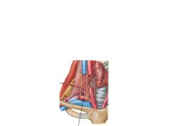
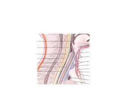
What muscle attachment courses between the R sublcavian a and v?
|

anterior scalene
|
|
|
How is the anatomy set up such that it's easier to hit the subclavian v than it is the subclavian artery? Where does the subclavian a become the axillary a?
|
The anterior scalene m lies between the anterior subclavian a and posterior subclavian a. The subclavian a become axillary when it reaches the lateral aspect of the 1st rib.
|
|
|
Draw the arteries coming off the subclavian a and into the neck. Recall the mnemonics for C-STAI-V and Sunday At Leff's Fans Open Pabsts & Munch Snacks
|
Draw the arteries coming off the subclavian a and into the neck. Recall the mnemonics for C-STAI-V and Sunday At Leff's Fans Open Pabsts & Munch Snacks
|