Reading...
![]()
Play button
![]()
Play button
![]()
Use LEFT and RIGHT arrow keys to navigate between flashcards;
Use UP and DOWN arrow keys to flip the card;
H to show hint;
A reads text to speech;
79 Cards in this Set
- Front
- Back
|
What is the origin of the Limb Mesenchyme?
|
Somatic Mesoderm (in other words, its just an extension of the body wall)
|
|
|
What is the limb primordia covered with?
|
Surface ectoderm
|
|
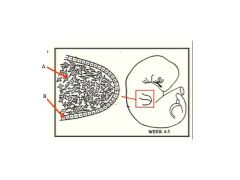
Label A and B
|

A = Somatic Mesoderm/Somitic Mesoderm
B = Surface Ectoderm |
|
|
How are the fully developed upper and lower limbs attached to the trunk?
|
Via pectoral and pelvic girdles, respectively
|
|
|
Establishment of the limb "field" occurs early in week:
|
4
|
|
|
Limb Fields arise from this tissue:
|
Somatic Mesoderm
|
|
|
Axial positioning is regulated by combinatorial expression of Hox genes. As a result, specific transcription factors are expressed in each LIMB FIELD. This TF if expressed in the upper limb and these TFs are expressed in the lower limb
|
Upper: Tbx5
Lower: Tbx4 and Pitx-1 |
|
|
When do LIMB BUDS begin to appear? (in days)
|
Upper limb = 26-27
Lower limb = 28-29 |
|
|
Limb buds seem to appear when somatic mesoderm cells begin to produce this grow factor:
|
Fgf-10
|
|
|
Budding is an inherent property of this tissue:
It is independent of this tissue: |
Mesoderm (independent of ectoderm)
|
|
|
Other limb precursor cells include: (there are 2; what do these tissues develop into):
|
1. Somitic Mesoderm (somite derived) forms Skeletal Muscles
2. Neural Crest forms Schwann cells and Spinal nn. |
|
|
What is AER and what induces it?
|
Apical Ectoderm Ridge is a thickened ridge of ectoderm that forms along the interface bn the dorsal and ventral surfaces of the limb bud. It is induced by production of Fgf-10 by mesenchyme cells
|
|

What type of tissue is A pointing to?
What type of tissue is B pointing to and what is it derived from? Note the dense region above the red arrow...what is that and what will it do? |
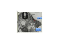
A = Surface Ectoderm
B = Mesenchyme (from somatic mesoderm) It's a myotome of a somite that will migrate into the limb bud. This invasion of Somitic cells into the Mesenchyme will form Skeletal Muscle |
|
|
Will the limb buds form without any signal from the Surface Ectoderm?
|
Yes - this is a unique feature of limb development
|
|

What are the arrows pointing to?
|
AER - Apical Ectodermal Ridge
|
|
|
After establishment of the limb field (phase 1) and budding (phase 2), when does step 3 happen? What is the defining change here?
|
Week 5-9 - elongation of the limb
|
|
|
During week 5, this forms and is the earliest indication of a hand/foot
|
A flat paddle-shaped plate develops at the distal end of the elongating limb.
|
|
|
By week 6, what can you see in the UL and LL?
|
Three segments and digital rays.
|
|
|
By week 8 what's going on with the UL and LL digital rays?
|
They are completely separate
|
|
|
What step also begins at step 3?
|
Step 4! Tissue formation and organization.
|
|
|
What happens during step 4 - tissue formation and organization.
|
The mesenchyme precursors begin to differentiate into tissues that you find in the limb.
|
|
|
In the central part of the limb, what condenses to form what?
|
Somatic MESOderm condenses to form cartilage and ultimately replaced by bone.
|
|
|
How does the skeletal muscles form?
|
Somite derived myoblasts migrate into the limb to form skeletal muscles.
|
|
|
How do the nerve and vascular precursors develop in the limb?
|
Motor axons and spinal nerves migrate into the limb following guidance cues from cells at the base of the limb bud.
|
|
|
Guidance cues for muscle and nerve development are received by:
|
Somatic mesoderm cells at the base of the limb bud.
|
|
|
Limb development occurs along three axes - describe what forms in each axis:
1. Proximal-Distal: 2. Anterior-Posterior: 3. Dorsal-Ventral: |
1. Elongation, segment formation
2. Digit development; cranial-caudal/preaxial-postaxial borders 3. Muscles and neurovascular structure compartments |
|
|
What happens whenever you remove the AER during development?
|
Elongation stops
|
|
|
Development along the proximal-distal axis of limb depends on:
|
Inductive interactions between the AER and underlying somatic mesoderm.
|
|
|
What happens if an additional AER is grafted onto the limb?
|
Duplication of limb structures occurs beginning from the time the graft is put in place.
|
|
|
What happens if mesoderm other than limb bud mesoderm is placed adjacent to the AER? What does this suggest?
|
Limb development is truncated...suggesting that only limb mesoderm is competent to respond to the signals from AER.
|
|

What can cause this?
|
Duplication of the AER
|
|

What can cause this?
|
Removal of the AER.
|
|
|
If mesoderm from the lower limb is grafted adjacent to AER in the upper limb, what happens? What does this suggest? (see p5)
|
The grafted tissue develops as though it were still in its original location. This suggests that the limb bud mesoderm is programmed as to the axial level at which it is located. For example, a transplanted wing AER will form a wing if placed near a leg.
|
|
|
What interaction is responsible for limb elongation? What is the source of the signals inducing elongation?
|
Interaction between Epithelial-Mesenchymal cells (AKA surface ectoderm and Somatic mesoderm, I think)
AER is the source of signals |
|
|
What four conclusions were made from the graft experiments?
|
1. Epi-mesenchymal interactions are responsible for limb elongation
2. AER is source of signals for limb elongation 3. Only limb mesenchyme (from somatic mesoderm) is competent of responding to its inductive signals 4. Axial and segmental patterning is inherent to the limb mesoderm |
|
|
What did studies on embryos with a "limbless" mutation suggest?
|
That somatic mesoderm produces limb bud independent of influence from ectoderm, but cannot sustain limb development further without AER-mesoderm interaction.
|
|
|
What TF does somatic mesoderm produce to induce formation of the AER?
What TF does the AER produce to maintain Fgf-10 production in the somatic mesoderm? What does ZPA produce |
1. Somatic mesoderm makes: Fgf-10
2. AER makes Fgf-8, 4, & 2 to maintain Fgf-10 in SM 3. ZPA produces Shh to maintain Fgf-8 secretion by AER |
|
|
What is the fate of the mesenchyme adjacent to the AER? (2 things)
|
-Forms segment specific skeletal elements
-Forms fibrous CT |
|
|
Summarize Model 1 vs Model 2 of limb development (p6)
|
1: AER signals diffuse along limb as it elongates. Mesenchyme leaving influence first become proximal limb, those 2nd become intermediate segment, those closest to AER become distal segment.
2. Segments along proximal-distal axis is specified early in limb development and AER signals are required for segments to expand and develop. |
|
|
What part of the arm does the limb bud mesoderm form?
1. Bone-wise 2. Fibrous connective tissue-wise |
1. Limb bones, clavicle, scapula.
2. Fibrous connective tissue that surrounds muscles and subdivides limbs into compartments and becomes the superficial fascia and dermis of the skin. |
|
|
What veins run along the preaxial border in UP and LL?
|
UP = cephalic vein
LL = great saphenous vein |
|
|
What tissue influences patterning along the anterior-posterior axis of the limb? What is this zone called?
|
Somatic mesoderm-derived mesenchymal cells along the posterior border of the limb - known as the Zone of Polarizing Activity.
|
|
|
What does the ZPA secrete?
|
Shh and Retinoic Acid
|
|
|
What does RA and Shh signaling induce?
|
Induces asymmetric, nested expression of Hox genes that mediates pattering for digit location along the anterior (thumb/big toe)-posterior (pinky/little toe) axis of the limb bud
|
|
|
The big digit forms on the ? border and the small digit forms on the ? border
|
Big digit: pre-axial
Small digit: post-axial |
|
|
High concentrations of SHH and RA would result in formation of which digit?
|
Small - in other words, this is closest to the ZPA
|
|
|
Describe where the AER remains once apoptosis begins to break down the digital rays
|
AER remains at the tips until the correct length and number of phalanges is achieved.
|
|
|
What structures are influenced by patterning along the dorsal-ventral axis?
|
1. Skeletal muscles
2. Neural and vascular structures 3. Limb compartmentalization |
|
|
What are the dorsal signals, where are they expressed, and what/where do they induce expression of? (p8)
|
Wnts and radical fringe (rFng) are expressed in Dorsal Ectoderm. They induce expression of Lmx-1 in dorsal mesenchyme.
|
|
|
What is the ventral signal, where is it expressed, and what does it inhibit? (p8)
|
Engrailed-1 (En-1) is the ventral signal expressed in the ventral ectoderm and it inhibits Lmx-1 in the ventral mesenchyme.
|
|
|
When do the limb buds begin to rotate?
|
Week 7
|
|
|
UL dermatome pattern:
LL dermatome pattern: |
UL: C3-T2
LL: L2-S3 |
|
|
Myoblasts forming the skeletal muscles within the limb bud as well as associated with pectoral and pelvic girdles are derived from:
|
Somitic (somite) mesoderm (ventral part of myotomes)
|
|
|
Patterning of limb muscles occur as specific axial levels - what are they?
|
C5-T1 for upper limb
L4-S3 for lower limb |
|
|
What stimulates myoblasts to migrate into the bud?
|
Signals from mesenchyme at the base of the limb bud.
|
|
|
Once myoblasts enter the limb bud, they organize into dorsal and ventral premuscle masses with respect to:
|
forming skeletal elements
|
|
|
Muscle patterning signals are believed to be derived from:
Splitting of muscles masses into specific muscles may be influenced by: |
Somatic mesoderm that forms the fibrous connective tissues that will be associated with the various muscle groups. Splitting of muscle masses may be influenced by signals directing blood vessel patterning.
|
|
|
When do spinal nerves grow into limb buds?
|
During week 5
|
|
|
Where do the guidance cues that spinal nerves follow into the limb bunds originate? (p9)
|
From mesenchyme cells at the base of the limb buds.
|
|
|
In what order do sensory and motor fibers enter limb buds? (what about NCC and what do they form?)
|
Motor fibers first then sensory fibers. Note: NCC also follow 2nd to form Schwann cells and melanoblasts.
|
|
|
What forms the supporting tissue for neurons and the melatonin in limbs>?
|
Schwann cells and melatonin are formed by Neural crest cells
|
|
|
How are the limb buds vascularized? (p9) What about the distal limb segment?
|
Sprouts from intersegmental arteries at specific axial levels enter the limb buds and form a vascular plexus of endothelial-lined tubes. Distally, an avascular zone forms under the ectoderm until all digits have formed.
|
|
|
When does limb rotation occur and about which axis does this occur?
|
Week 7 - around the proximal-distal axis
|
|
|
What forms dermis vs epidermis of skin?
|
1. Dermis = somatic mesoderm
2. Epidermis = Ectoderm |
|
|
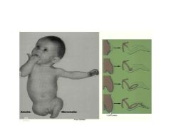
Failure of Formation of Limb Parts is subdivided into two categories:
|
1. Transverse Type (Amelia or Meromelia)
2. Longitudinal Type (Meromelia) |
|
|
Define transverse type malformation:
|
Truncation/amputation anywhere along the proximal-distal axis. Eg missing hand, foot, etc.
|
|
|
Define Longitudinal Type malformation:
|
Absense of limb parts occuring along the anterior-posterior axis. Eg. missing radius = anterior absence
Missing ulna, pinky = posterior absence |
|
|
Deformity with too few digits in the middle is called (i.e. missing digits 2, 3, or 4):
|
Oligodactyly (oligo = not enough). AKA "Lobster Claw"
|
|
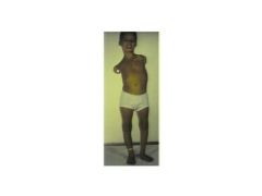
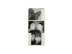
What is this called? What is it caused by? Is it longitudinal or transverse anomaly?
|

Phocomelia. Possibly by thalidomide. This is a longitudinal anomaly
|
|
|
Define Phocomelia
|
Defect where distal segment of the limb is attached to a more proximal segment with the intervening segment missing or directly attached to the girdle.
|
|

Anomaly on L? R? Caused by failure of: Specifically, the problem on the R might also be caused by:
|
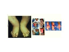
Syndactyly and Sirenomelia
Caused by interdigital apoptosis. The R might also be caused by abnormal tailbud development. |
|

How might this happen>
|
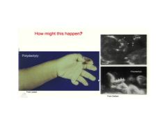
Over activity of Zone of Polarizing Activity.
|
|

What is the condition on the L and R called?
|
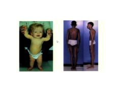
L Achondroplasia
R Gigantism (Marfan Syndrome) |
|

What is this condition called? What is wrong?
|
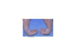
Talipes Equinovarus AKA Clubbed Foot. The talus bone is missing/abnormal (varus = turned in)
|
|
|
Define Defect vs Distortion
|
Using clubbed foot as an example: Defect would be if the Talas bone is missing. Distortion would be if the structure is all there but the foot/leg is misshappen bc of pressure throughout development in the womb.
|
|
|
Term used to describe a shortened digit due to lack of certain joints?
Termed used to describe long thin digits. |
1. Brachydactyly
2. Arachnodactyly |
|
|
Developmental Dysplasia of the Hip (congenital dislocation of the hip) is caused by (3):
|
1. Underdevelopment of the femoral head and/or hip socket
2. Maternal hormones that sofent connective tissue 3. Position of the uterus 4. Sometimes there is a hereditary component |
|
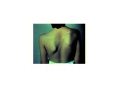
What is this?
|

Sprengel Deformity - elevated scapula
|
|
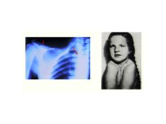
What is this? In mice, they can induce this by creating a heterozygote mouse defective in this gene:
|

Cleidocranial Dysplasia. RunX2 (aka CBFA1)
|