![]()
![]()
![]()
Use LEFT and RIGHT arrow keys to navigate between flashcards;
Use UP and DOWN arrow keys to flip the card;
H to show hint;
A reads text to speech;
26 Cards in this Set
- Front
- Back
|
Phases of haemostasis
|
1. Vascular
2. Platelet 3. Coagulation |
|
|
Vascular phase: vasospasm
|
Cutting the wall of a blood vessels, triggers smooth muscle contraction.
- Vasospasm, lasting around 30 mins to slow or even stop the flow of blood |
|
|
Vascular phase: endothelium
|
- Endothelial cells contract and expose underlying BM to blood stream
- Endothelial cells release local factors and hormones e.g. ADP, tissue factor, prostacyclin, endothelins - Endothelial plasma membranes become "sticky", facilitating the attachment of platelets and even sticking to the opposite endothelial wall |
|
|
Endothelins
|
Peptide hormones that:
- stimulate smooth muscle contraction and promote vascular spasm - stimulate the division of endothelial cells, smooth muscle cells and fibroblasts to accelerate the repair process |
|
|
Platelet phase
|
Begins with the attachment of platelets to endothelial surfaces, BM and exposed collagen fibres
Platelet adhesion > platetlet aggregation > platelet plug |
|
|
Activation of platelets
|
First sign of activation: become more spherical, develop cytoplasmic processes to link with other platelets. Release a variety of compounds:
1. ADP stimulates platelet aggregation and secretion 2. Thromoxane A2 and serotonin stimulate vascular spasm 3. Clotting factors are proteins 4. PDGF is a peptide that promotes vessel repair 5. Calcium ions are required for platelet aggregation and in the clotting process |
|
|
Progression of platelet phase
|
ADP, thomboxane and calcium are released from each arriving platelet, stimulating further aggregation
- this is a positive feedback loop |
|
|
Role of prostacyclin
|
A prostaglandin that inhibits platelet aggregation and is released by endothelial cells. Controls and restricts platelet aggregation to the injury site.
- This is reinforced by inhibitory compounds released by WBCs |
|
|
Role of plasma enzyme
|
Circulating plasma enzymes break down ADP near the plug, restricting the response
|
|
|
Inhibition of plug formation
|
- Serotonin blocks the action of ADP at high concentrations
- Circulating plasma enzymes - Inhibitory WBC compounds - Prostacyclin inhibits platelet aggregation - development of a blood clot isolates the plug |
|
|
Coagulation phase
|
A complex sequence of steps leading to the conversion of circulating fibrinogen into the insoluble protein fibrin.
- As the fibrin network grows, blood cells and additional platelets are trapped in the fibrous tangle, forming a blood clot. |
|
|
TImings of phases
|
Vascular and platelet phases begin with a few seconds of injury
Coagulation does not start until 30 seconds later |
|
|
Clotting factors
|
- Normal clotting depends on procoagulants in the blood: Ca2+ and 11 proteins
- Many of the proteins are proenzymes, which are converted to active enzymes and direct essential reactions in the clotting response |
|
|
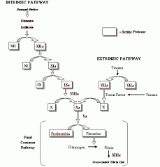
Pathways initiating clotting cascade
|
Extrinsic: begins outside the blood stream in the vessel wall (appropriate activation)
Intrinsic: begins in the blood vessel with the activation of a circulating proenzyme These two pathways converge at the common pathway |
|
|
Draw clottting cascade
|

|
|
|
Extrinsic pathway
|
Appropriate activation:
- TF/FIII is released by damaged endothelial cells or peripheral tissues - The more damage, the more TF released |
|
|
Intrinsic pathway
|
Inappropriate activation:
- activation of proenzymes exposed to collagen at the site of insult |
|
|
The common pathway
|
Enzymes from either pathway activate FX, forming the enzyme prothrombinase.
- Prothrombinase converts prothrombin into thrombin - Thrombin completes the clotting cascade by converting fibrinogen to insoluble fibrin |
|
|
Clot retraction
|
One the fibrin mesh-work has formed, platelets and RBCs stick to the fibrin strands.
- The platelets then contract, and the entire clot undergoes clot retraction - This process continues over 30-60mins |
|
|
Feedback control of coagulation
|
Thrombin generated in the common pathway stimulates clotting by:
- Stimulating the formation of TF 2. Stimulating the release od PF-3 by platelets This is a positive feedback loop, stimulating intrinsic and extrinsic pathways |
|
|
Restriction of coagulation: antithrombin-III
|
- Normal plasma includes several anticoagulants e.g. antithrombin-III inhibits several factors, including thrombin
- Heparin is released by basophils and mast cells, and accelerates the activation of antithrombin-III |
|
|
Restriction of coagulation: aspirin
|
- Aspirin inhibits the production of thombozane-A2 and prostaglandins.
- Prevents platelet aggregation and so clot formation - Prolongs bleeding time |
|
|
Restriction of coagulation: thrombomodulin
|
- Thombomodulin is released by endothelial cells and binds to thrombin
- Activated thrombin then activates protein C - Protein C is a plasma protein that inactivates several clotting factors and stimulates the formation of plasmin - Plasmin gradually breaks down fibrin strands |
|
|
Restriction of coagulation: prostacyclin
|
Prostacyclin released during the platelet phase inhibits platelet aggregation and opposes the stimulatory action of thrombin and ADP
|
|
|
Restriction of coagulation: Vitamin K
|
Sufficient vitamin K is needed in the liver to synthesise 4 of the clotting factors
- long term lack of Vit K causes breakdown of the common pathway and eventual destruction of the clotting cascade |
|
|
Fibrinolysis
|
Activation of proenzyme plasminogen by thrombin and tissue plasminogen activator (t-PA) released by damaged tissues
- produces the enzyme plasmin which begins digesting the clot |

