![]()
![]()
![]()
Use LEFT and RIGHT arrow keys to navigate between flashcards;
Use UP and DOWN arrow keys to flip the card;
H to show hint;
A reads text to speech;
68 Cards in this Set
- Front
- Back
|
The base of the Heart faces __1__ and to the __2__
|
1. Superiorly
2. Right |
|
|
Apex points __1__ and __2__
|
Left and Down
|
|
|
Base consists primarily of the ____
|
Left Atrium, part of RA and Proximal part of Great Vessels
|
|
|
The Apex consists of the _____
|
left ventricle
|
|
|
Female heart Weight = 1
Male weight = 2 |
female = 250-300 g
male = 300-350 |
|
|
Right side of heart contains this type of blood
Left contains this kind of blood |
Right = venous
Left = Arterial |
|
|
Tricuspid and Bicuspid valves each has something that the Pulmonary and Aortic Valves don't have. What is it?
|
Papillary Muscles
*all have Chordae Tendinae |
|
|
Layer of Heart that has fat in it
|
Myocardium
|
|
|
2 classifications of Endocardium
|
1. Mural Endocardium = lining the cardiac chambers
2. Valvular = covering the valves |
|
|
_______arrangement of myocytes is essential for the transmission of electrical impulses through the myocardium, and a synchronized contraction of cardiac chambers
|
Syncytial
|
|
|
2 surfaces of Pericardial Sac
|
Epicardium = lining the heart itself
Pericardium = surface facing the Epicardium |
|
|
Occur at the ends of myocytes and maintain cell to cell cohesion
|
Intercalated Disks
|
|
|
Present at the intercalated disks and are low-resistence paths between cells that allow for rapid electrical spread of action potentials
|
Gap Junctions
|
|
|
Contractile unit of a Myocardial cell
|
Sarcomere
|
|
|
Thick filaments of Sarcomere
Thin filaments of Sarcomere |
Myosin
Actin |
|
|
Separates sarcomeres
|
Z line
|
|
|
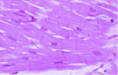
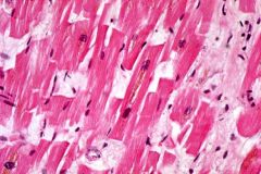
Position of Myocardial nuclei
|

centrally located
|
|
|
Most common disturbance caused by MI
|
Electrical disturbance
|
|
|
Cell membrane of Myocardiocyte
|
Sarcolemma
|
|
|
Site of storage and release of Ca+ for excitation-contraction coupling
|
Sarcoplasmic Reticulum
|
|
|
Strength of contraction depends on...
|
initial length of cardiac myocytes
*max strength = 2.2 microns, anything longer causes weaker contractions |
|
|
Troponin that is the Calcium binding regulatory protein
|
Troponin C
|
|
|
What do blood vessels in the Valves usually represent?
|
Angiogenesis due to BACTERIAL INFECTION = Valvular Endocarditis
*usually Valves contain no blood vessels |
|
|
Effects of aging on the Heart Chambers
|
Left Atrial DILATION
Left Ventricle cavity reduction |
|
|
Effects of aging on Valves
|
1. Dystrophic Calcification
2. Fibrosis 3. focal thickenings |
|
|
Effect of aging on Coronary Arteries
|
Atherosclerosis
|
|
|
4 effects of aging on the Myocardium
|

Hypertrophy
Brown Atrophy Basophilic Degeneration Amyloidosis |
|
|
Explain Senile Amyloidosis
|
Amyloid is derived from proteins in blood -> gets deposited in myocardium but not removed -> builds up so much that myocytes can't contract/conduct well
|
|
|
3 Effects of aging on the Aorta
|
Dilation and Elongation
Atherosclerosis Elastic fragmentation |
|
|
5 Mechanisms of Heart Failure
*What is most common? |
1. Pump Failure****
2. Flow obstruction 3. Regurgitation of blood 4. Electric conduction disorders 5. rupture of circulatory system |
|
|
are cJUN and cFOS oncogenic or Tumor suppressors?
|
Oncogenic
|
|
|
Those with Hypertrophic Myocytes have these genes activated
|
Embryonic genes
-Beta-myosin heavy chain -Skeletal alpha-actin *allow for increased muscle activity |
|
|
Consequences of Hypertrophy
|

increased demand for O2 -> relative ischemia -> death of myocytes -> fibrosis -> weakness of myocardium
|
|
|
What is Forward Heart Failure?
|
ischemia due to reduced Systolic output
|
|
|
What is Backward Failure?
|
congestion due to inadequate emptying of the heart chambers (stagnation of blood)
|
|
|
4 causes of Left Sided Heart Failure
|
1. Ischemic Heart Disease
2. Hypertension 3. Aortic/Mitral valve diseases 4. Myocarditis |
|
|
Left Atrial Dilation due to LV failure can result in these 3 things (sequential)
|
Atrial Fibrilation -> blood is shoved about and can coagulate to form Thrombi -> Thrombi detach and form Emboli
|
|
|
2 syndromes that may have Congenital Heart Disease
|
Down Syndrome
DiGeorge Syndrome |
|
|
Environmental causes of CHD's
|
Rubella
Alcohol |
|
|
Most likely cause of CHD's
|
Multifactorial effects of several genes, maternal, and environmental factors
|
|
|
Most common CHD
|
Ventricular Septal Defect (VSD)
|
|
|
Most common Cyanotic CHD
|
Tetralogy of Fallot
|
|
|
Most common type of Atrial Septal Defect
|
ASD secundum (ASD 2)
|
|
|
ASD 2 is a defect in the _______
|
Foramen Ovale
|
|
|
ASD Primum is adjacent to the _______ and is associated with deformities of the _____
|
AV valves
Mitral valve |
|
|
ASD Sinus Venosus is located at the entry level of the __1__ into the __2__
|
1. Superior Vena Cava
2. Right Atrium |
|
|
ASD-1 results embryonically from a defect in ______ development
|
Endocardial Cushion (associated with Down Syndrome)
|
|
|
Ventricular Septal Defect usually involves this part of ventricle
|
Membranous part of Septum (90%)
*muscular part = 10% |
|
|
Large VSD's are usually in this part of ventricle
|
Membranous
*small in Muscular part |
|
|
Explain VSD
|
1. Left to Right shunt at first
2. With increased blood going from LV to RV to Lungs, Pulmonary HTN develops 3. Pulmonary HTN results in late Right to Left shunt 4. Mixing of RV blood into LV = LATE CYANOSIS |
|
|
Reversal of the shunt from L-R to R-L is called?
|
Eisenmenger Syndrome
|
|
|
What is the Ductus Arteriosus?
|
In fetus allows venous blood to bypass the lungs (R->L shunt)
RV -> Pulmonary Artery -> Aorta |
|
|
Patent Ductus Arteriosus is associated with _______
|
Congenital Rubella Syndrome
|
|
|
This closes PDA
This keeps PDA open |
Indomethacin
PGE2 |
|
|
PDA:
If the shunt reverses, unoxygenated blood enters the Aorta below the ______ resulting in Cyanosis in the _______ |
Subclavian Artery
Lower extremities |
|
|
PDA presents with this type of murmur
|
Machine-like
|
|
|
Tetralogy of Fallot pathogenesis
|
PROVe
1. Pulmonary Stenosis 2. RVH 3. Overiding Aorta 4. VSD |
|
|
Tetralogy of Fallot Symptoms depend on the severity of _________
|
Pulmonary Stenosis
|
|
|
T-of-F:
-Severe stenosis increases __1__ shunting and causes __2__ |
1. Right to Left
2. Cyanosis |
|
|
Tetralogy of Fallot:
-Cyanosis occurs: 1 -compensatory __2__ -__3__ __4__ |
1. soon after birth
2. polycythemia (high RBC's) 3. Clubbing of fingers/toes 4. Retarded growth |
|
|
Explain Transposition of Great Vessels
|
Aorta receives blood from right side of heart
Lungs receive blood from left side of heart |
|
|
What is required for compatibility of life with Transposition?
|
VSD
or PDA |
|
|
In Transposition, which has greater compatibility with life?
|
VSD
|
|
|
Two forms of Coarctation of the Aorta
|
Infantile
Adult |
|
|
Explain Infantile Coartation of Aorta
|
- Aortic arch and Ascending Aorta are markedly narrowed
- Ductus Arteriosus turns into PDA -PDA serves as conduit for blood flow below the narrowing -Head and Upper extremities are hypoxic -Lower extremeties are Cyanotic -High Mortality RATE |
|
|
Explain Adult Coarctation of Aorta
|
-narrowing is opposite the Ductus Arteriosus which closes
-Hypertension in Upper Extremities and Head -Notching of Ribs -Low pressure in the Legs -Symptomatic in ADULTHOOD |
|
|
CHD's that cause Early Cyanosis (Blue Babies)
|
3 T's:
Tetralogy of Fallot Transposition of Vessels Truncus Arteriosus |
|
|
Infantile Coarctation of the Aorta is associated with.....
|
Turner's Syndrome
|

