Reading...
![]()
Play button
![]()
Play button
![]()
Use LEFT and RIGHT arrow keys to navigate between flashcards;
Use UP and DOWN arrow keys to flip the card;
H to show hint;
A reads text to speech;
33 Cards in this Set
- Front
- Back
|
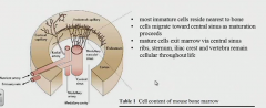
The primary or ___ lymphoid tissues are in the ___ and ___ and are the sites of immune cell ___
|
central
bone marrow and thymus immune cell development |
|
|
The secondary or ___ lymphoid tissues are in what tissues/organs. These are the sites of .... (?)
|
peripheral
spleen, lymph nodes, mucosal-associated tissue mature cell residence and initiation of adaptive immune responses |
|
|
Hematopoiesis is _____
most immature cells reside ____ to bone they mature in _____ as they start to be matured they migrate toward ___ and exit the marrow via ___ |
compartmentalized
nearest islands of maturation central sinus central sinus |
|
|
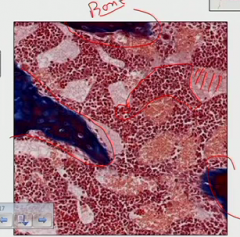
Which section of the bone does the hematopoiesis occur in?
In this area there's a network of ___ and ___ cells which are the sources of ___ and ___ |
The spongy bones toward the end. It creates 3D environment (whatever that means)
reticular and endothelial cells for growth factors and cytokines (drive proliferation and differentiation) |
|
|
The non-immune structural cells in the spongy bone have to ___ with the hematopoietic cells in order for them to ___
|
physically interact for them to mature
|
|
|
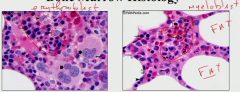
When you compare different cells in the marrow, which do you see at highest concentrations
|
Cells going to granulocytes-> neutrophils, eosinophils, and basophils
Followed by lymphocytes and RBC's |
|
ID some key features on here-- what's happening where?
|
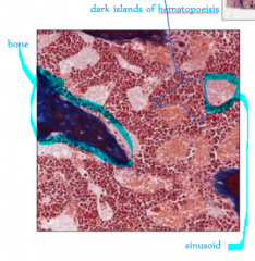
bone trabeculae make pockets for the developing immune cells.
The dark regions: islands of hematopoiesis where they're developing The lighter regions: sinusoids where the cells go, making their way to the central sinus |
|
What are we looking at, and how is it relevant to the hematopoietic story?
|
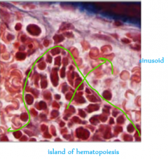
Darker is hematopoietic part; lighter is sinusoid. They are separated by a layer of epithelium which the migrating cells have to get through similar to extravasation in inflammation
|
|
|
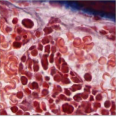
How do you ID a neutrophil?
|
multilobulated nucleus
|
|
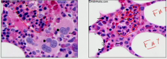
compare the two circled areas
|
can tell by the nuclei mainly
|
|
|
it is super duper important that the reticular cell infrastructure ___ developing immune cells to ...
The reticular cells can be referred to as ___ The reticular cells can communicate with progenitor cells via what mechanisms? |
physically contacts to drive maturation (he's said this 3 times already)
stromal cells receptor-receptor or by releasing or showing (still attached) cytokines to interact with progenitor's receptors |
|
|
When would we use the stromal-progenitor interactions clinically?
Name a drug and its descriptor |
If pt is neutropenic (low levels making them susceptible to infection)
leukine (it's a GM-CSF: granulocyte monocyte- colony stimulation factor) |
|
|
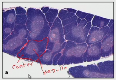
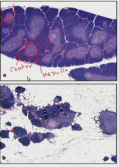
Gross description of thymus and its location
It has ___ ___ within lobes, each consisting of an outer ___ and inner ___ |
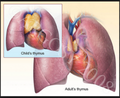
bi-lobed structure under sternum
(before this year started, I had always pictured the thymus being up in the neck-- I didn't realize how far down in the chest it was) multiple lobules (like a functional unit) with a (darker) cortex and (lighter) medulla |
|
|
Where does the maturation of T cells occur within the thymus? What do you call the cells that left the bone marrow to go to the thymus to do this?
So what do the other parts do? |
thymocytes come in to do thymopoiesis development into mature T cells in the cortex of the lobules.
The cortical epithelial cells provide cell contact signals and growth factors |
|
|
Mature T cells leave the lobule and enter the blood via ...
|
venules in corticomedullary junction
|
|
|
As you get older your thymus shrivels up as fat replaces the parenchymal tissue which is really called _______ which causes ___
|
age-related thymic involution
"immunosenescence"- there's still some of the tissue remaining but the functioning capacity decreases |
|

Which area is all the thymocytes going to mature t cells?
Just like in the bone marrow, the maturing cells migrate to ___ portions of the ___ and then go out. The ___ contains many macrophages which ... |
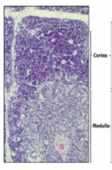
cortex
lower portions of the cortex medulla; they clean up debris |
|
|
At the top of the lobule cortex, the cells are dividing super rapidly but more than __% of them will die. They are trying to ___(generally described)____ but it's a very ___ process
|
90%
trying to make an antigen receptor but it's a very inefficient process that makes a lot of mistakes (also explains all the debris the macrophages in the medulla are eating) |
|
|
In more detail outline what the thymocyte goes into (what is on its surface) and decide to become either __ or __. Once they make that choice that put another molecule on their surface depending on their choice-- what is on each respective cell?
|
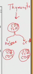
CD3 and TCR (t cell receptor which recognizes antigen)
helper cell adds CD4 CTL (cytotoxic lymphocyte) adds CD8 |
|
|
In the secondary lymphoid tissues, it's all about the mature immune cells....
|
getting initiated. Before they were maturing.
|
|
|
The spleen is a __ bed that takes out ___, and is the site of ___ immune response to ___ antigens. The spleen has ___ which penetrate the organ carrying ___ which break down into ___. They are encased in ___ which is called the ___
|
Filtration bed taking out debris, and the site of adaptive immune response to blood-borne antigens. It has fibrous trabeculae which carry blood vessels which break down into central arterioles which are encased in T lymphocytes as the periarteriolar lymphoid sheath (PALS).
|
|
|
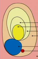
From the PALS there are outpockets called ___ which are rich in ___. There are two types of these-- name them, what the difference is, and how you can see it histologically.
|
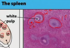
lymphoid follicles rich in B cells.
primary lymphoid follicle - resting B cells (just purple), no active immune response secondary lymphoid follicle - outer mantle of resting B cells (purple ring) with a central germinal center containing proliferating B cells (white center). (In the picture, he's marked the red CA = central arteriole surrounded by PALS and then off to the right circled again is a secondary lymphoid follicle) |
|
Breaking down a secondary lymphoid follicle: looking at this, do you know what the different areas are and what they contain?
|
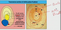
The marginal zone has B cells resting and is darker. If this were a primary lymphoid follicle, everything inside its boundaries would be dark. This is secondary, so the granular center is present lighter in color with active B cells. CA central arteriole and PALS towards bottom. PALS has T cells.
|
|
|
Process of circulating Ag into the spleen-- they come in to the spleen in the ___ via the ___. Most of them will course right past the follicle and dump into the ___ which ____ them. The parts that go into the follicle get dumped into the ___. There the lymphocytes try to find their Ag. If they do find it, they ___.
|
blood via central arteriole.
dump into the red pulp which just eats all the debris including the Ag perifollicular zone If they find the Ag they migrate-- depending if they are B/T they go different places. |
|
|
Lymph nodes are the sites of ___ immune response to ___ antigens. The lymph enters the node via ___ that are kind of at the head of the node. The cells get in via ___. Within the node, there are areas rich in B and T cells and there can be also be ___ just like in the spleen which differentiate primary and secondary follicles.
|
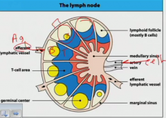
tissue-borne
afferent lymphatic vessel the blood supply germinal center (he circled three spots- furthest right is a primary; the other two secondary). |
|
|
The main difference between how the spleen and lymph nodes work is...
|
how the Ag goes in. The system inside is basically the same.
|
|
|
In the lymph: what is a subcapsular sinus? What's in it?
|
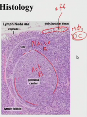
The afferent lymphatics thereby the antigens get dumped into it. It contains a lot of macrophage and dendritic cells (which are responsible for presenting antigen to T cells)
|
|
|
In the medulla of the lymph node, it is rich in...
|
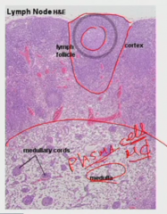
macrophages and plasma cells.
The plasma cells can sit here comfortably and pump out antibodies. |
|
|
Where do the cells extravasate from the vessels to go into the lymph node?
|
postcapillary high endothelial venule
|
|
|
Mucosal-associated lymphoid tissue is referring to what?
|
Under the epithelium of all mucosal tissue sites are lymph node like tissue. It has immune cells- both effector and naive cells.
|
|
|
How do Ag get into the mucosal-associated lymphoid tissue?
|
Just come from the lumen of the organ.
|
|
|
There are diffuse immune cells in this region just below the epithelium.
|
lamina propria
|
|
|
In the ileum, there are regions loaded with lymphoid follicles. These are called what and what do they do? How does the Ag get in?
|
Peyer's patches
The Ag comes in through the top from the lumen via M cells, a special epithelium cell. It puts the Ag in and dendritic cells take them to start the immune response. |