![]()
![]()
![]()
Use LEFT and RIGHT arrow keys to navigate between flashcards;
Use UP and DOWN arrow keys to flip the card;
H to show hint;
A reads text to speech;
157 Cards in this Set
- Front
- Back
|
The ___ provides a case for the brain, brainstem, meninges, cranial nerves, and blood vessels The cranium has a roof called the ___ and a floor that is called the __ which consist of what 5 bones? |
cranial vault calvaria cranial base ethmoid bone, sphenoid bones, parts of the temporal, occipital, and frontal bones |
|
|
The cranium in ADULTS is formed by a series of which eight bones ? In adults the bones are joined by ___ In newborns the cranial bones are separated by areas of the ___ how many fontanelles are there? |
frontal bone, pair of parietal and temporal bones, occipital, sphenoid and ethmoid bone fontanelles 6 ( 1 ant, 1 post, 2 pairs of sphenoidal fontanelles, and 2 pairs of mastoid fontanelles) |
|
|
The skull is oriented so that the inferior margin of the orbit and the superior margin of the external acoustic meatus (auditory canal) of both sides lie in the same horizontal plane called the ___ The __ is the weakest part of the skull, it is located at the juntion of what 4 bones? The pterion is located where? |
frankfort horizontal/ auriculo-orbital plane pterion frontal, temporal, parietal and sphenoid bones the temporal fossa (or temple) |
|
|
The pterion bone is two fingers breadths superior to the ___ and a thumb breadth posterior to the ___ of the frontal bone. this is an important landmark on the lateral aspect of the skull because the ___ lies in the groove on the internal aspect of the lateral wall of the skull |
zygomatic arch zygomatic process anterior branch of the middle meningeal artery (maxillary artery) |
|
|
Injury to the ___ (during a boxing match) may lacerate the skin and cause profuse bleeding Bruising of the skin around the eye causes fluid and blood to accumulate in the surrounding connective tissue, which gravitates into the __ eyelid |
superciliary arches superior (black eye) |
|
|
Fractures of the Calvaria (cranium): __ fractures are fragments of bone are depressed inward to compress or injure the brain _ are the most frequent types of fractures usually occurring at the point of impact. these fractures lines often radiate away in two directions __ is a fracture where the bone is broken in several fragments |
depressed linear skull comminuted |
|
|
laceration of the middle meningeal artery results in ___ laceration of the cerebral artery results in __ laceration f the cerebral vein (bridging veins) results in __ (time delay) subarachnoid hemorrhage usually results from what? |
epidural hematoma subarachnoid hemorrhage dural border hematoma (subdural) hypertension or ruptured aneurysm |
|
|
if the area of the skull is thick at the site of impact, the bone usually does what? In a ___ fracture, no fracture occurs at the site of impact but on occurs on the opposite site of the skull |
bends inward without fracturing countercoup/counterblow |
|
|
The cranial cavity contains what? (4) *What does the anterior cranial fossa contain? (4) |
-the brain and its meninges, cranial nerves, arteries and veins, venous sinuses -frontal lobes of the brain, crista galli for attachment of the falx cerebrum, cribiform plate for the olfactory nerve, foramen cecum for emissary veins from the nasal mucosa to the dural sinuses of the brain (superior sagittal sinus)
|
|
|
What does the middle cranial fossa contains? (10)
*the superior orbital fissure contains CN 3, 4, 5(1), and 6, and the superior ophthalmic vein. the optic canal contains CN 2, and ophthalmic artery)* |
Temporal lobes, optic canal, superior orbital fissure, foramen rotundum (V2), foramen ovale (V3), foramen spinosum (middle meningeal artery and vein), foramen lacerum (fat), carotid canal (ICA, internal carotid nerve plexus), sella turcica (pituitary gland), and groove for the middle meningeal artery (temporal bone opposite the pterion) |
|
|
What does the posterior cranial fossa contain? (8)
*foramen magnum contains the medulla oblongata, vertebral artery, and meningeal arteries* |
cerebellum, pons and medulla oblongata, tentorium cerebelli, foramen magnum, hypoglossal canal (CN 12), internal auditory meatus (CN 7, and 8), and the condylar canal and groove for the transverse sinus |
|
|
Which veins drain into the dural venous sinuses? *The superior sagittal sinus receives blood from the what? (4) *this sinus lies along the borders of the falx cerebri* |
vertebral venous plexus from the pelvis, emissary veins of the scalp and the nasal mucosa, angular vein via the superior ophthalmic vein form the face and nose (this drains retrogradely into the cavity sinus) -nasal cavity, scalp, meningeal vein and superior cerebral vein, *(occipital sinus as well)* |
|
|
The __ occupies the posterior two thirds of the free edge of the falx cerebri. The inferior sagittal sinus joins the __ to form the straight sinus which empties in the confluence of sinuses where is the confluence of sinus located? |
inferior sagittal sinus great cerebral vein internal occipital protuberance |
|
|
The __ begins at the internal occipital protuberance and it becomes the sigmoid sinus, which after exiting the jugular foramen it becomes the internal jugular vein The _ drains the cavernous sinus into the transverse sinus The _ exits the jugular foramen and then it drains into the jugular vein and it also drains the cavernous sinus |
transverse sinus superior petrosal sinus inferior petrosall sinus |
|
|
*in the cavernous sinus the __ and the ___ runs through the sinus (or horizontally), and CN _, _,_ and _ run lateral to it (or vertically from superior to inferior)* *which veins drain into the cavernous sinus?* (3) |
internal carotid artery and CN 6 -CN 3, 4, 5(1) and 5(2) -superior and inferior ophthalmic viens and the central vein of the retina
*5(2)= maxillary nerve 5(1)=olfactory nerve the internal carotid artery goes through the internal carotid canal* |
|
|
_ artery is a branch of the subclavian artery and it passes through the transverse foramina of C6-C1 to enter the foramen magnum of the skull. _ vein begins inside of the skull and passes through the foramina of C1-C7 to drain into the subclavian vein |
vertebral vertebral |
|
|
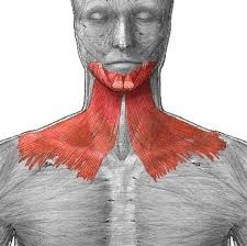
The subcutaneous tissue of the neck contains nerves and superficial vessels which are covered by a flat muscle that ascends on the face from the anterior aspect of the neck called the __ Laryngeal Prominence also called Adams apple: thyroid cartiage, is located at __ Hyoid Bone is superior to the thyroid cartilage located at ___, inferior to the chin in the median plane |
platysma C4/5 C3 |
|
|
Cricoid Cartilage inferior to the thyroid cartilage at the level of __ ____ fossa: a triangular depression located posterior to the clavicular head of the SCM. This is the pressure point for the subclavian artery |
C6 supraclavicular |
|
name the muscle, origin and insertion, innervation, and function |
Platysma fascia and skin over the pectoralis major and deltoid muscles inferior border of the mandible and skin of the lower face cervical branch of CN 7 muscle of facial expression |
|

|
Sternocleidomastoid - Sternal head- from the manubrium, Clavicular head- superior surface of the medial 1/3 of the clavicle -lateral surface of the mastoid process of the temporal bone and superior nuchal line of the occipital bone -CN11 and branch of C2 and C3 tilts the head to the lateral side and superiorly turn it to the opposite side. They flex the neck (both muscles)
|
|

|
Trapezius Accessory Spinal Nerve (CN 11) and cervical plexus (C3/4) -elevates and rotates the scapula upwards in arm elevation, its lower portion depresses and rotates the scapula downwards (adducts the scapula, tilts its chin, draws back acromion, rotates the scapula) |
|
|
The lateral aspect of the neck is called the ___ and is divided into the anterior and posterior cervical triangles *In the posterior cervical triangle the __ artery passes over the apex of the triangle before it ascends over the posterior aspect of the head* *what is the floor of the of the posterior cervical triangle?* |
cervical triangles occipital (from superior to inferior)- splenius capitus, levator scapulae, scalenus posterior, medius, anterior muscles |
|
|
*The posterior cervical triangle is subdivided into two triangles by what?* *What are those two triangles?* *What are the contents of the posterior cervical triangle?* (12) |
-Inferior belly of the omohyoid muscle -Occipital, and supraclavicular/omoclavicular/subclavian triangles -External jugular vein, subclavian vein, Third portion of the subclavian artery, transverse cervical artery (thryocervical trunk), superificial cervical (transverse cervical) artery, suprascapular artery (thyrocervical trunk), Occipital artery (ECA), CN 11, brachial plexus, cervical plexus, anterior cervical and superficial cervical Lymph nodesc |
|
|
*What forms the external the jugular vein?* *The retromandibular vein is formed inside the parotid gland by the union of the _ and _ *The brachial plexus lies between what two muscles?* |
-By the posterior division of the retromandibular vein and the posterior auricular veins -superior temporal vein and maxillary vein -scalene anterior and scalene medius |
|
|
*Where is the cervical plexus (ventral rami of C1-4) located?* What is the boundries of the posterior cervical triangle? |
-deep to the internal jugular vein and SCM -Anterior: posterior border of the SCM, posterior: anterior border of the trapezius. Inferior: clavicle |
|
|
What are the nerves of the Cervical Plexus (sensory branches)? (4) _ supplies the skin over the lateral side of the occipital region *_ accompanies the external jugular vein and supplies the skin over the angle of the mandible* _supplies the skin over the anterior triangle of the neck __ nerve supplies the skin of the anterior aspect of the chest and shoulder. *The medial and lateral supraclavicular nerves supply which joints respectively?* |
lesser occipital (C2), Greater Auricular (C2/3), and transverse cutaneous Nerve/colli (C2/3) and supraclavicular Nerve C3/4 -lesser occipital nerve -great auricular nerve -transverse cervical cutaneous -supraclavicular nerve -sternoclavicular and acromioclavicular joints |
|
|
What he muscular branches of the cervical Plexus? (2) *_ is the loop that arises from the cervical plexus and supplies what muscle?* *Where is the anacervicalis nerve embedded?* |
Phrenic Nerve (C3/4/5) and the Ana cercvicalis Anacervicalis -infrahyoid muscle -the carotid sheath (in the fascia) **but it is not a content of the sheath** |
|
|
What is the boundaries of the anterior cervical triangle? What are the contents of the anterior cervical triangle? What is the hyoid bone connected to? Muscles of the anterior cervical triangle are referred to as __ which are responsible for steadying or moving the hyoid bone and larynx (these are muscles are the suprahyiod and infrahyiod muscles) |
Anterior: median line of the neck, posterior: anterior border of the SCM, Superior: inferior border of the mandible -muscles, veins, arteries, nerves, LN and viscera -mandible, manubrium of the sternum and scapula -hyiod muscles |
|
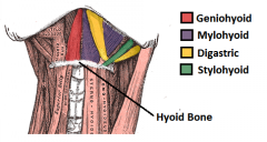
which are the suprahyiod muscles? |
mylohyiod, geniohyiod, stylohyiod, and diagastric muscles |
|
|
__ form the floor of the mouth, innervated by CN 7 _ reinforce the floor of the mouth. innervated by CN 12 __ is innvervated by CN 7 |
Mylohyiod muscle Geniohyiod muscle stylohyiod muscle |
|
|
__ is a strap like muscle that has two bellies (anterior and posterior) that descends toward the hyiod bone. The two bellies are joined by a common central tendon called the ___ that is connected to the body of the hyiod bone The anterior belly originates from the __ and the posterior originates from the __ What is the innervation of the anterior and posterior belly respectively? |
digastric muscles intermediate tendon fossa of the mandible mastoid process nerve to mylohyiod muscle (CN 5) and the CN 7 |
|
|
What are the infrahyiod muscles (strap muscles)? What is the function of the infrahyiod muscles? __ and _ is innervated by the ansa cervicalis (C1, C2, C3) __ is responsible for closing the laryngeal opening during swallowing and preventing food from entering the larynx. This muscles is innervated by what nerve?
|
-sternohyiod, sternothyroid, thyrohyiod, and omohyiod muscles -they anchor and depress the hyiod bone and the larynx during swallowing and speaking -sternohyiod and sternothryoid muscles -thryohyiod musle -C1 fibers with CN 12 |
|
|
__ muscle has two bellies joined by an intermediate tendon. The inferior attachment is the superior border of the scapula and the superior attachment is the inferior aspect of the hyiod muscle. Innervated by the ansa cervicalis |
Omohyiod muscle |
|
|
What two muscles divided the anterior cervical traingle into four smaller regions? What are the four smaller triangular subdivisions of the anterior cervical traingle? |
-superior belly of the omohyiod muscle and diagastric muscle -Diagastric triangle (submandibular), submental triangle (suprahyiod), Carotid triangle, and muscular triangle |
|
|
*What does the carotid sheath contain? (5)* What is the main artery of the head and neck? The __ begins at the bifurcation of the brachiocephalic trunk, posterior to the right sternoclavicular joint The __ artery arises from the aortic arch and ascends into the neck posterior to the left sternoclavicular joint |
Internal jugular vein, Deep cervical lymph nodes, vagus nerve, Common Carotid, and internal carotid arteries *ansa cervicalis is embedded in it* -RCC -LCC |
|
|
*Each carotid artery ascends within the carotid sheath to the level of the __ where it terminates by dividing into the internal and external carotid arteries* What arteries have no branches in the neck? |
thyroid cartilage common carotid and internal carotid arteries |
|
|
what are the branches of the external carotid artery? (8) |
Superior thyroid, ascending pharyngeal, lingual, facial, occipital, posterior auricular, superficial temporal (end artery) and maxillary (end artery) arteries |
|
|
Carotid sinus is a small dilation of the __ part of the ICA. It is blood pressure regulating area innervated by the __, _ and _. Carotid sinus responds to changes in what? The Carotid Sinus is a __ that responds to increase the arterial blood pressure by slowing the heart rate with vagus nerve |
proximal CN 9, 10 and sympathetic divisions of ANS arterial blood pressure baroreceptor |
|
|
The __ is a small ovoid mass of tissue located at the bifurcation of the common carotid arteries, in close relation with the carotid sinus The carotid body is a __ that responds to changes in the chemical composition of the blood. It responds to increase CO2 or H+, or decrease O2 or pH which all leads to an increase in __ The carotid body is innervated by the what? |
carotid body chemoreceptor respiration Carotid sinus nerve (branch of CN 9), vagus nerve (CN X), and sympathetic nerves |
|
|
What are the layers of the cervical viscera? (3) What is in the superficial endocrine layer? (3) What is in the middle respiratory layer? (2) What is in the deep alimentary layer? (2) |
Superificial endocrine, middle respiratory, and deep alimentary -thymus, thyroid gland, parathyroid -larynx, trachea -pharynx, esophagus |
|
|
The superficial layer of the cervical fascia ensheathes the __ muscle *The superficial layer of the DEEP cervical fascia splits at the __ and covers the __ laterally and the __ medially** it envelopes what?* *the superficial layer of the deep cervical fascia forms what?* |
-platysma -mandible -masseter -medial surface of the medial pterygoid -SCM, trapezius, submandibular, and parotid glands -forms the floor of submandibular space |
|
|
*The middle layer of the Deep cervical fascia envelopes what?* (5) *What are the borders of the muscular division in the middle layer of the deep cervical fascia?* *The muscular division envelopes what?* |
-thyroid, trachea, esophagus, pharynx, larynx -superior: hyiod and thyroid cartilage; inferior: sternum, clavicle and scapula -infrahyiod strap muscles |
|
|
*What are the two layers of the deep layer of the cervical fascia?* *What are the borders of the Alar layer?* *what are the borders of the prevertebral layer?* *The prevertebral layer envelopes the __ and __. it extends laterally as the _ sheath |
-alar and prevertebral layer -superior border: skull base; inferior border: upper mediastinum at T1/2 -Superior border: skull base; inferior border: coccyx -vertebral bodies and deep muscles of the neck -axillary sheath |
|
|
he scalp consist of five layers of tissue that cover the osseous cranium. It extends from the ___ on the posterior aspect of the skull to the __ anteriorly. Laterally it extends to the level of the __
What are the layers of the scalp? (5) |
superior nuchal line supraorbital margins zygomatic arches -skin, subcutaneous tissue with large superficial vessels and nerves, epicranial aponeurosis, Loose areolar CT, ad pericranium (periostium) |
|
|
The epicranial aponeurosis (galea aponerotica) is also called what? ___ with emissary veins, is clinically important because superficial infection of the scalp may spread to deeper layers through the __ veins which communicate wit the __ sinus |
subaponeurotic tissue/ loose areolar CT emissary veins intracranial (subdural) venous |
|
|
What three layers of the scalp are referred to as the scalp proper and are clinically and surgically regarded as one single layer? |
-skin, subcutaneous tissue, and the epicranial aponeurosis |
|
|
The trigeminal nerve is made up of what three nerves? The ophthalmic nerve consist of what 5 nerves? The Maxillary nerve consist of what 3 nerves? The Mandibular nerve consist of what 3 nerves? |
- (V1) Ophthalmic (superior orbital fissure), (V2) Maxillary (foramen rotandum), V(3)- Mandibular (foramen ovale) -supraorbital*, supratrochlear*, external nasal, infratrochlear, lacrimal nerves -zygomaticofacial N*, infraorbital, zygomaticotemperol nerves -buccal, auriculotemperol*, mental nerves |
|
|
What are the branches of the external carotid artery? Which artery is the terminal branch of the external carotid artery and accompanies the auricotemperol nerve? What are the branches of the internal carotid artery? |
occipital, posterior auricular, and superficial temperol artery -superficial temporal artery -supratrochlear and supraoribital arteries. |
|
|
The cutaneous, sensory supply of the of the face is largely through what nerve? *The __ nerve from the cervical plexus (C2) contributes to the innervation of the face by innervating the skin over the angle of the mandible and the anterior/posterior auricle* The motor innervation is supplied by the __ nerve and its branches to the superficial muscles of the neck (platysma), muscles of facial expression and the muscles of the scalp |
Trigeminal nerve and its divisions -great auricular nerve -facial nerve (CN 7)
|
|
|
The sensory component of the facial nerve supplies taste to the anterior 2/3 of the __ and the it also conveys general sensation from the __, and it is a secretory motor (parasympathetic) to thesubmandibular, sublingual, intralingual salivary glands (submandibular ganglion), and lacrimal gland (pterygopalatine ganglion) |
tongue external acoustic meatus |
|
|
The facial nerve emerges from the skull through the ___ between the mastoid and styloid processes of temporal bone. It enters the __ gland in the parotid region of the face and it divides into five terminal branchess |
stylomastoid foramen parotid gland |
|
|
What are the five main branches of the facial nerve? what do they innervate respectively? What are the other branches of the facial nerve and their innervations? (3) |
1. temporal -frontalis and orbicularis oculi muscles 2. Zygomatic- orbicularis oculi muscle 3. Buccal- muscles of the upper lip and nostrils 4. mandibular -muscles of the lower lip 5. Cervical- platysma muscle -posterior auricular (occipitalis muscle and auricles), Nerve to stapedius (middle ear), nerve to post. belly of diagastric and stylohyoid muscles |
|
|
Sensitivity to sound: __ (hyperacusis) taste to the anterior 2/3 of tongue: sweet, sour, bitter (_) What are 4 symptoms of damage to the facial nerve? *presentation is dependent on where damage occurs (facial canal vs geniculate ganglion vs stylomastoid foramen vs cerebellopontine angle)* |
audiometer chorda tympani -ipsilateral muscle paralysis (bells Palsy), loss of taste to the anterior 2/3 of the tongue, hyperacusis, decreased lacrimation |
|
|
In the facial nerve damage, when the patient is asked to smile. the facial musculature pulls to the opposite side of the injury. When the patient attempts to close his/her eye, the eyeball rolls upwards and outwards aka ___ What are the arteries of the face? (3) |
bells phenomenon -facial artery (ECA), superficial temporal, transverse facial artery (from the superficial temporal) |
|
|
*_ artery runs over the lower border of the mandible and it anastomoses with the branches of the ophthalmic artery (ICA). This point is a connection between the internal and external carotid arteries.* What are the branches of the facial artery? (2) *Of the branches is the terminal branch which anastomoses with the dorsal nasal and palpebral arteries of the ophthalmic artery (ICA)* |
Facial artery -inferior labial artery, superior labial artery and the angular artery -angular artery
|
|
|
the facial vein is formed by what? *The facial vein, at the inferior margin of the mandible it is joined by the __ vein and it terminates into the __ vein. it communicates with the __ vein which drains into the _ sinus of the skull which the drains into the __ vein* |
union of the supratrochlear and subraorbital veins near the medial angle of the eye (medial anthus) (begins as the angular vein) -anterior retromandibular -internal jugular vein -superior ophthalmic vein -cavernous -internal jugular |
|
|
The muscle of the face are innervated by what nerve?
What are the muscle of the scalp and their functions? (2) What are the muscles of the nose? (2) |
-CN 7 (facial nerve) -frontalis/occipitofrontalis (elevates the eyebrows/suprised face); wrinkles the forehead/frown) and the procerus muscle (continuation of the frontalis muscle/pulls the eyebrows down) -transverse (compressor naris) and the alar (dilator naris) |
|
|
What are the muscles of the orbit? (2) __ closes the eyelid, dilates the lacrimal glands. paralysis of this muscle results in inability to close the eye and drooping of the lower eyelid (ectropia) and spilling of the tears (epiphora) _ innervated by CN 3 and is the muscle of the eyelid
|
-orbicularis oculi and levator palpebrae -orbicularis oculi -levator palpebrae - |
|
|
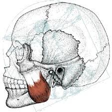
Muscles of mastication are innervated by the trigeminal nerve ___ What are the four muscles of mastication? |
-CN (V3)/ mandibular -masseter, temporalis, lateral pterygoid, medial pterygoid |
|
name the muscle, origin, insertion, innervation, and function |
-masseter o: zygomatic arch and maxilla I: angle and lateral surface ramus I: mandibular F: elevates the mandible and also assists in protraction, retraction and side to side motion |
|

|
temporalis O: temporal line of the parietal bones I: coronoid process I: mandibular F: elevates the mandible with its vertical fibers, retracts the mandible with its horizontal fibers, and lateral movement during chewing |
|

|
lateral pterygoid muscle O:great wing of the sphenoid, lateral surface of the lateral pterygoid plate I: condyle of the mandible I: mandibular F: protrudes the mandible and lateral movements during chewing |
|

|
Media; pterygoid muscle O: medial side of lateral pterygoid plate I: angle of the mandible I: mandibular F: raises the mandible (adducts) |
|
|
The temporomandibular joint is a __ joint located between the glenoid cavity of the temporal bone and the condyle of the mandible. What is the innervation of the temporalmanidbular joint?
|
-synovial -auriculotemporal nerve (V3), maseteric and deep temporal nerves |
|
|
__ ligament prevents posterior and lateral displacement of the mandible __ ligament limits distention of the mandible in an inferior direction. Most commonly damaged in an inferior alveolar nerve block __ ligament limits excess opening _ is the fibrous capsule |
temporomandibular (lateral) -sphenomandibular stylomandibular capsule ligament |
|
|
What are the movements of the of the temporomandibular joints? (3) __ is when the mandible is moved left/right -non- working/balancing side __ is when the mandible is moved left/right- working side |
rotation (opening and closing the mouth), translation (protrusion and retrusion of the mandible) and grinding movements during mastication (mediotrusive, laterotrusive) -mediotrusive -laterotrusive |
|
|
TMJ disorder is dysfunction of the _ joint? What are sign and symptoms? (7) treament? (5) |
temporomanidbular joint -periorbital headache, difficulty chewing, moving the jaw (open/close) with associated pain, tinnitus in the ear (fullness, ringing), clicking and popping of the jaw with possible dislocation or subluxation, otitis without infection, dizziness, and cervical pain -NSAID'S, orthodontic splints, habit change (diet), stress reduction, surgery |
|
|
Fractures of the mandible are classified as favorable or unfavorable. A __ fracture is one that is held in apposition and alignment to the natural pull of the attached muscles. A __ fractures are those that are displaced by the pull of attached muscles |
favorable unfavorable |
|
|
After diagnosing a fracture, it is important to identify any pathological dental condition in which the fracture lies. A fracture that extends into a __ may result in delayed healing and osteomyelitis What are signs and symptons of mandibular fractures? Treatments? |
periapical abscess -pain, welling, bruising, sublingual hematoma, trismus (muscle spasm), mental anesthesia, occlusal disharmony palpable, condylar palpable in fossa, bleeding, and other infra and intra oral features (airway obstruction can occur due to hematoma formation or edema) *bilateral fractures result in higher risk for airway obstruction* -pain relief and assesment for airway obstruction, surgical fixation , prevention of infection |
|
|
Fractures of the mandible include the __ (single and uncommon 2%), the __ (often transverse, associated with TMJ dislocation on the same side 30%), the __ (which is oblique and may involve the 3rd molar 25%), and the __ (usually pass through a canine tooth 25%) |
coronoid process neck of mandible angle of mandible body of mandible
|
|
|
__ is a method of demonstrating virtually all mandibular fractures, including the coronoid and condylar processes. But sometimes symphysis menti fractures may not been seen The OPG must be supplemented with a _ to establish displacement of fractures. this will demonstrate the body and symphysis menti and may also provide tangential veiws of the rami and t he necks of the condyles |
Orthopantomogram (OPG)/panorex posterioanterior (PA) mandible |
|
|
In the absence of an OPG, the __ will demonstrate the body and ramus on each side, the articulation at the TMJ may also be assessed __ fractures are more rare than mandibular fractures, usually associated with the nose, teeth fractures. Causes a great deal of swelling so delayed repair is not common, CTs are used to evaluate the fracture, and surgical reduction with open fixation is usually required for treatment |
lateral oblique radiographs maxillary fractures (three types: le fort 1, 2, and 3) |
|
|
The parotid region is made out of what? (3) The parotid region lies below the what? The parotid region is a space between what? *The parotid duct aka __ measures approximately 5cm in length, runs along the __ muscle and it turns sharply to pierce the __ muscle. It enters the oral cavity opposite of the crown of the __ tooth* |
parotid gland, ramus of the mandible (anteriorly) and the styloid process ( medially) -zygomatic arch -mastoid process & the neck and the ramus of the mandible -stensens duct -masseter -buccinator -2nd maxillary molar tooth |
|
|
What are the structures that lie within the parotid gland from lateral to medial? (3) Facial nerve has two branches, __ to the posterior belly of the diagastric and stylohyoid muscles and the __ nerve The retromandibular vein is formed by what? What is the blood supply of the parotid gland? Innervation of parotid gland? __ produces a thin watery salivary flow, __ stimulates a thick mucos salivary discharge |
Facial nerve, retromandibular vein, ECA, parotid LN -muscular branches -posterior auricular nerve -the union of the maxillary and superficial temporal veins within the parotid glands -ECA Auriculotemporal nerve (V3), Glossopharyngeal (CN 9 parasympathetic) , and sympathetic cervical ganglion -CN 9 -sympathetic ganglion |
|
|
The submandibular region is bounded by what? What nerves are in the submandibular region? (3) blood vessels? (4) What is the function of the digastric muscle?
|
inferior border of the mandible superiorly and the two bellies of the digastric muscle -lingual nerve, CN 9, and hypoglossal CN 11 -facial artery and vein, lingual artery and vein -depresses the mandible and elevated the hyoid |
|
name the muscle, origin, insertion, innervation and function |
-hyoglossus muscle O: hyoid bone I: side of the tongee N: CN 12 F: depresses the tongue
|
|

|
-Genioglossus O: superior mental spine I: tip and middle of the tongue N: CN 12 F: protrude the tongue |
|

|
-Styloglossus O: tip of the styloid process **I: side of the tongue passes between the middle and superior constrictors of the pharynx** N: CN 12 F: draws the tongue up and back |
|
|
The submandibular gland is a salivary gland that lies in the __ triangle of the neck. The submandibular duct aka __ opens in the oral cavity in the ___ at the sides of the frenulum of the tonuge Innervation: Parasymthetic (__ preganglionic w/chorda tympani to the _ ganglion) and Sympathetic from the __ ganglion |
digastric whartons duct sublingual papillae CN 7 submandibular cervical |
|
|
The sublingual gland is innervated by the __ nerve. The sublingual nerve lies in the floor of the mouth between the __ and __ The lingual nerve carries the general sensation from the anterior 2/3 of the __, it is a branch of the __ nerve (division of the trigeminal nerve). It is found on the lateral surface of the __ muscle and crossess the __ twice |
lingual nerve (V3) (has parasympathetic and sympathetic fibers) mandible genioglossus muscles -tongue -mandibular nerve -hyloglossus submandibular duct
|
|
|
What are the boundries of the temporal boundries ? What are the contents of the temporal region? (5) |
anteriorly: superior temporal line, anteriorly: frontal process of the zygomatic bone, inferiorly: zygomatic arch -temporalis muscle, deep temporal nerve (V3), deep temporal artery (maxillary artery), auriculotemporal nerve, and superficial temporal artery |
|
|
The _ region is a space located below the temporal region and the behind the maxilla What are the boundries of the infratemporal fossa? |
infratemporal Superior: infratemporal surface of the sphenoid bone, anterior: posterior surface of the maxilla and inferior orbital fissure, medial : lateral pterygoid muscle and pterygomaxillay fissure , lateral: ramus and coronoid process of the mandible |
|
|
What are the contents of the infratemporal fossa? (8) What the branches of the maxillary artery? (4) |
-lower border of the temporalis muscle, lateral and medial pterygoid muscle, pterygoid venous plexus, maxillary artery and its branches, maxillary nerve, mandibular nerve, otic ganglion -inferior aveolar, middle meningeal, branches to the external auditory meatus and tympanic membrane, branches to the muscle of mastication |
|
|
What does the infratemporal fossa communicate with? (4) |
-temporal fossa, inferior orbital fissure, middle cranial fossa w/ foramen ovale and foramen spinosum, and the mandibular canal with the mandibular foramen |
|
|
The pterygopalatine fossa is a major distributing center for the branches of the __ nerve and artery. It is the area inferior to the __ between the pterygoid process of the sphenoid bone, posteriorly, and the posterior aspect of the __ anteriorly What are the boundries? What are the contents? (3)
|
maxillary apex of the orbit maxilla -Superior (body of the sphenoid bone), anterior (posterior surface of the maxilla), posterior (lateral pterygoid plate), medial (perpendicular plate of the palatine bone) -Maxillary nerve and artery, pterygopalatine ganglion |
|
|
The maxillary nerve leaves the skull through the __ reaching the pterygopalatine fossa. It then goes through the __ where it changes names to the __ nerve. The infraorbital nerve exits through the infraorbital foramen to become __ to the face (it is the end nerve) What are the branches of the maxillary nerve?(4) |
foramen rotundum infraorbital fissure infraorbital sensory - meningeal , communicating branches to the pterygopalatine ganglion, superior alveolar nerves, zygomatic nerve |
|
|
__ is a terminal branch of the ECA. *Branches: __ mucous membrane of nasal cavity and septum. it is the end artery of the maxillary* __ is a venous network that surrounds the lateral pterygoid muscle. It is located partly between the temporalis and lateral pterygoid muscles and partly between the two pterygoid muscles .It communicates with what? *__ joins the superficial temporal vein in the parotid gland to form the retromandibular vein* |
Maxillary sphenopalatine -pterygoid venous plexus -facial vein w/ the deep facial vein -maxillary vein |
|
|
pterygoid ganglion (parasympathetic) lies below the _ nerve in front of the __ canal and behind the __. It maybe blocked through the __ notch |
maxillary nerve pterygoid middle nasal concha mandibular
|
|
|
What is the roof of the orbit? (2) What is the lateral wall of the orbit? (3) What is the medial wall of the orbit? (3) What is the floor of the orbit? (3) |
lesser wing of sphenoid bone, frontal bone -zygomatic process of frontal bone, greater wing of the frontal bone, orbital surface of zygomatic bone -frontal process of maxilla, lacrimal bine, lateral mass of ethmoid bone -perpendicular plate of palatine bone, orbital surface of maxilla, zygomatic bone |
|
|
Indirect trauma that displaces the orbital walls is called a __ fracture fractures of the medial wall may affect the _ and _ sinuses, whereas fractures of the inferior wall may affect the _ sinus Fracture in the __ (part of the orbit) can result in objects entering the frontal lobe of the brain |
blowout sphenoidal, and ethmoidal maxillary superior orbit wall |
|
|
Orbital fractures often results in intraorbital bleeding, which may increase the pressure and push on the eyeball causing a condition known as _ __ is when there is direct trauma to the periorbital region often causes swelling hemorrhage into the eyelids and extravasation of blood into the periorbital skin, often happens in contact sports and boxing |
exophthalmus periorbital ecchyymosis |
|
|
The place where the eyelids meet is called the what? What is the normal width of the palpbral fissure? The eyelids are moveable folds covered by thin skin externally and internally by what? What is the palpebral conjunctiva is reflected onto the eyeball where it is continuous with ___ __ covers the sclera and is adherent to the periphery of the cornea it also contains small blood vessels |
angle of the eye (canthus)/medial and lateral canthi 9mm (6-10) -palebebral conjunctiva -bulbar conjunctiva
|
|
|
What maintains the shape and rigidity of the superior and inferior eyelids? What is the skeleton of the eyelids? The tarsal plates are suspended in the eyelids by the __, a connective tissue membrane that spans from the tarsal plates to the margins of the orbit where it is continuous with the periosteum.
|
tarsal plate tarsal plate orbital septum |
|
|
__ glands are embedded in the tarsal plates and produce an oily secretion which lubricates the edges of the eyelids and prevents the from sticking together when the eyelids are closed. This secretion also from a barrier to prevent the over flow of tears. The Cillary (of zeis) glands are large sebaceous glands associated with the eye lashes that secrete what into the eyelash hair? What does this prevent? |
-Meibomian (tarsal) -sebum -from the eyelash from becoming to brittle
|
|
|
_ glands are modified sweet glands near the lid margins, empties several places including same route as glands of route as glands of Zeis. Their function is unknown What ligament is between the nose and the medial canthus of the eye and connects the tarsal plates to the medial margin of the orbit? *What muscle originates and inserts onto the medial palpebral ligament?* |
-Glands of Moll -medial palpebral ligament -orbicularis oculi |
|
|
__ ligaments attaches the tarsal plates to the lateral margin of the orbit but *DOES NOT* provide for muscle attachment |
lateral palpebral ligament |
|
Name the muscle and function. Where does it insert to? |
levator palpebrae superioris elevates the eyelid inserts into the superior tarsal plate with the orbicularis oculi |
|
|
__ is an involuntary smooth muscle innervated by sympathetic fibers. Assists in elevation of the eyelid and inserts in the superior tarsal plate _ muscle is thought to depress the inferior eyelid, and widen the palpebral fissure __ closes the eyelids __ is the sensory innervation to the upper eyelid _ is the sensory innervation to the lower eyelid _ innervates the orbicularis oculi muscle |
-superior tarsal muscle -inferior tarsal muscle -orbicularis oculi muscle -CN 5 (V1) ophthalmic -CN 5 (V2) maxillary -CN 7 |
|
|
__ innervates the levator palpebrae superioris muscle *sympathetic use postganglionic fibers from the superior cervical ganglion to superior tarsal muscle* If the ducts of the ciliary glands (of zies) become obstructed, a painful, suppurative swelling a _ develops on the eyelids |
CN 3 sty (hordeolum) |
|
|
Obstruction of the tarsal gland produces an inflammation referred to as __ that may protrude toward the eyeball and rub against the cornea __ is manifested by bright or dark red patches deep to and in the bulbar conjunctiva. these may result from inflammation or injury |
Chalazion subconjunctival hemorrhages |
|
|
A direct blow to the eye, excessive hard blowing of the nose, and *__* of coughing or violent sneezing may cause hemorrhages resulting from rupture of the small sunconjunctival capillaries Lesion of CN __ will cause paralysis of the __ muscle causing ptsosis (drooping) of the upper eyelid |
paroxysms 3/occulomotor levator palpebrae superioris |
|
|
Damage to CN _ will cause paralysis of the __ muscle resulting in the inability to close the eyelids. The normal rapid blinking protective reflex is also lost The loss of muscle the lower eyelid causes the lid to sag (becomes everted) away from the surface f the eye. This leads to drying of the cornea resulting in irritation which leads to excessive but insufficient __ |
3/occulomotor orbicularis oculi lacrimation |
|
|
Loss of innervation to the orbicularis oculi by CN 7 causes the inability to voluntarily close the ___ tightly and the __ droops away resulting in spillage of tears aka __ The lacrimal sac is located at the medial margin of the oribit. The lacrimal sac is connected to the __ duct Secretory fibers to the lacrimal glands are from the ___ nerve (CN 7) and the nerve of the __ canal |
eyelids lower eyelids epiphora greater petrosal nerve pterygoid |
|
|
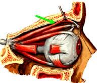
* Where does the nasolacrimal duct drain into?* Excessive lacrimation may result from obstruction of the __ duct What are the contents of the orbit? (5) What are the 3 layers of the eye? The fibrosus tunic layer contains what? (2) The vascular tunic layer contains what? (3) The neural tunic layer contains what? (1) |
inferior nasal meatus lacrimal eyeball and associated muscles, nerves, blood vessels, fat, and lacrimal apparatus -retina, choroid or middle (vascular) coat, external or fibrosus coat -sclera, cornea -iris, ciliary body, choroid -retina
|
|
|
The __ is a thin delicate membranous supporting structure covered externally by the __ and internally by the __ What is the blood supply of the retina? |
retina choroid vitrous body central artery a branch of the ophthalmic artery *(which runs through the optic disc)* |
|
|
What are the two layers of the retina? Contents of retina: 1)__ is where the optic nerve exits. it contains no photoreceptors (it is a blind spot). And the central retinal vessels radiate from here. The retina is most adherent here 2)The __ is a condition in which the disc is swollen and has a indistinct margins 3) the __ nerve which exits at the disc, is covered by the same meningeal layers that cover the brain, therefore increased pressure in the CSF can affect the disc |
outer pigment cell layer and inner neural layer (ends anteriorly at the posterior margin of the ciliary body) -optic papilla -papilledema -optic nerve |
|
|
__ can occur as a consequence of increased intracranial pressure secondary to hemorrhage or a tumor. Increased IOP can result in __ of the optic disc. *Normal IOP= 10-21 mmHg (avg=16 mmHg)* The __ is a small, oval, yellowish area located lateral to the optic papilla The __ is the central depressed part of the macula. *it is the area of the most acute vision* |
papilladema cupping macula lutea fovea centralis |
|
|
__ occurs when the outer pigmented and inner neural layers o the retina separate. Individuals who are ___ due to more elliptical eyeball, are more at risk for detachment because their retina is typically thinned or "stretched" more than that of the normal eye _ is the physical deterioration of the macula lutea, first symptom is loss of visual acuity |
retinal detachment near sighted -macular degeneration (ARMD- age related macular degeneration) |
|
|
In hypertensive retinopathy there are 3 classes: __ is arteriolar narrowing, tortuosities, irregular calibre __ arteriovenous nipping __ is flammed shaped and blot hemorrhages, cotton wool sits and hard exuadates __ is papilledema
|
Grade 1 Grade 2 Grade 3 Grade 4 |
|
|
_ is highly vascular dark layer located between the sclera and the retina. Capillaries into this layer supply deeper layer of the retina by __ _ is the anterior, thickened portion of the choroid The choroid is connected to the lens by the __ ligaments The _ is the processes folds on the internal surfaces that secrete aqueous humor, a watery fluid that fills the anterior and posterior chamber of the eye |
choroid diffusion ciliary body suspensory ciliary processes |
|
|
The ciliary body also contains the __ muscle which upon contraction allows the lens to bulge (accommodate) by relaxing the suspensory ligaments. *The ciliary muscle is a smooth muscle innervated by the parasympathetic nerves from the occulomotor nerve which synapse in the __ ganaglion* |
ciliary muscle ciliary ganglion |
|
|
Distant vision (no accommodation) is when the ciliary muscle is __ and the lens is _ because it is stretched by taut suspensory ligaments Near vision (accommodation) is when the ciliary muscle is __ lens is __ because of the tension on the lens is released. This is enhanced focus _ is age related inability to tread close up whereby the lens remains flattened in spite of relaxation of the suspensory ligaments by ciliary muscle contraction
|
relaxed flat contracted rounded Presbyopia |
|
|
The __ is the colored circular membrane that lies between the cornea and the lens. In the central opening the _ allows light to pass thorough The iris contains two smooth muscles, the _ dilates the pupil and is innervated by sympathetic nerves. The __ constricts the pupil and is innervated by the parasympathetic nerves |
iris pupil dilater pupillae sphincter pupillae |
|
|
__ is when small capacites within the lens, over time may coalesce to completely obscure the len. this is a major cause of blindness. this results from aging , diabetes, intraocular infections, excessive UV light, and glaucoma. The fibrosus coat is made up of what? (2) _ is the white, dense connective tissue portion _ is the anterior, transparent portion
|
Cataracts sclera and cornea sclera cornea |
|
|
The corneal reflex is a _ reflex. It elicits a bilateral response (touching one side results in blinking both eyes). the afferent limb is the _ nerve and the efferent is the __ nerve Chambers of the eye: the anterior cavity is anterior to the __ and posterior to the __. The anterior cavity is divided by the iris: The anterior chamber of the anterior cavity is between what? The posterior chamber of the anterior cavity is between what? |
protective CN 5 (trigeminal) CN 7 (facial) lens cornea iris and corn lens and iris
|
|
|
The anterior cavity contains __. What are the main functions of aqueous humor? The aqueous humor is secreted into the __, goes around the __, and down through the _ then into the __. It leaves the anterior chamber through the canal of __ to join the venous blood stream |
aqueous humor -it removes waste products and helps maintain the chemical environment within the anterior and posterior chambers of the eye -posterior chamber -lens then pupil -anterior chamber schlemm |
|
|
_ is a disease characterized an increased IOP over 22 mmHg, it results from increased production or diminshed reabsorption of aqueous humor. Fluid buildup causes a posterior dislocation of the lens and substantial increase in pressure in the posterior chamber _ humor fills the space between the lens and retina |
Glaucoma vitrous humor |
|
|
The muscles of the eyeball are innervated by what? (3) Movements of the superior oblique and inferior oblique muscle of the eye on the transverse axis will have what kind of movements? Movements along the A/P axis will have what kind of movements? |
-superior oblique (from CN 4), lateral rectus ( from CN 6) and R3 -elevation and depression -intorsion and extorsion |
|
|
On the transverse axis: elevation of the eye is done by the __, Depression of the eye is done by the __ and __. On the anteroposterior axis intorsion is done by the __ muscle and extorsion is done by the __ muscle. On the vertical axis abduction is done by the __ muscle, and adduction is done bu the _ muscle. Convergence is done by the __ muscle |
superior rectus and inferior oblique inferior rectus and superior oblique superior oblique inferior oblique lateral rectus medial rectus medial rectus
|
|
|
__ muscles are the best elevators/depressors when the eye is adducted _ muscles are the best elevators/depressors when the eye is abducted Therefore which muscles which muscle are responsible for eye movement in different positions of gaze? |
oblique rectus oblique and rectus |
|
|
Superior rectus will __ the eye, innervated by nerve? Inferior rectus will __ the eye, innervation? The inferior oblique will have __ on the eye, innervation? The medial rectus will _ the eye, innervation? the lateral rectus will _ the eye, innervation? The superior oblique will have __ on the eye, innervation? |
elevates, 3 depresses, 3 elevation and extorsion, 3 adducts (only), 3 abducts (only), 6 depression and intorsion, 4 |
|
|
___ will accompany the ophthalmic artery (ICA), it is a nerve of sight and its ends in the ___ What are the three main branches of the ophthalmic nerve (sensory branch of CN 5) ? Which of these branches supplies the skin f the lateral eyelid. What is the branches of the frontal branch? Which of the branches enters the orbit w/the superior orbital fissure? |
optic nerve optic chiasm lacrimal, frontal, nasocilliary lacrimal frontal nasocilliary |
|
|
Which CN nerve provides parasympathetic supply to constrictor pupillae and ciliary muscles w/ the ciliary ganglion? What muscles does CN 3 innervate preganglion ___----> postganglionic___----->___ |
CN 3 -levator palpebrae superioris, superior, medial, inferior rectus muscles, and inferior oblique muscles edinger westphal nucleus (viseral motor) ciliary ganglion constrictor pupillae |
|
|
The occulomotor nerve exits the skull with the _ fissure and divides into the superior and inferior division. Which division innervates the superior rectus and levator palpebrae muscle? Which division innervates the medial and inferior rectus and inferior oblique? Damage: Ptsosis is paralysis of the __ muscle, __ is when there is unopposed action of the lateral rectus and superior oblique muscles, mydrasis is paralysis of the _ muscle, inability to accommodate is called cycloplegia and is paralysis of the __ muscle |
superior orbital anterior inferior levator palpebrae lateral strabismus constrictor pupillae ciliary |
|
|
__ only supplies the superior oblique muscle, and it exits through the superior orbital fissure. *Damage: Pts with forth nerve palsies can obtain binocular vision by tilting their heads to the unaffected side, thereby causing the normal eye to __ and line up wit the extorted, affected eye* |
trochlear (CN 4) nerve intort *patients is asked to to look downward with the eye adducted. limitation of movement indicates paralysis or damage* |
|
|
The __ nerve innervates the lateral rectus muscle. __ happens when you have paralysis of the lateral rectus muscle. A result is diplopia (double vision). Head turned to side of lesion restores binocular vision Damage to the abducen nerve causes what? |
abducens (CN 6) nerve strabismus *(inability to direct both eyes towards the same object)* -inability to abduct the eye (medial strabismus) |
|
|
***Ciliary ganglion is at the back of the orbit it is lateral to the __ and medial to the ___. The occulomotor nerves gives _ fibers to the ciliary ganglion which then synapse with neurons in the ciliary ganglion and the _ fibers go to the ciliary muscle and sphincter pupillae by short ciliary nerves. Symathethic fibers from the __ hitch a ride with the _ artery to the ciliary ganlion WITHOUT synapsing and then by short ciliary nerves go the the dilator pupillae, and palpebral or tarsal muscles*** |
optic nerve lateral rectus preganglionic postganglionic SCG internal carotid |
|
|
Blood vessels of the orbit: __ artery travels in the optic canal inferior to the optic nerve What are the branches of the ophthalmic artery? The lacrimal artery, which gives the recurrent meningeal artery, will have anastomoses with which artery? The orbit is drained by which veins? *The superior ophthalmic vein joins the facial vein and drains into the __ sinus* |
-ophthalmic artery -central artery of the retina, posterior ciliary, supraorbital, supratrochlear, dorsal nasal, anterior and posterior ethmoidal, and lacrimal arteries -middle meningeal artery -superior and inferior ophthalmic -cavernous
|
|
|
The nose is divided into the right and left cavities by the what? The naris is divided into the _ area (superior 1/3), and the _ area (2/3) |
Nasal septum olfactory respiratory |
|
|
What are the functions of the nose? (5) The nostrils are bounded medially by the __ and laterally by the __ |
Respiration, Olfaction, filtration of dust, humidification of inspired air, reception of secretions from the paranasal sinuses and nasolacrimal ducts -nasal septum -ala of the nose |
|
|
What are the 3 components of the nose? * the nasal cavities extend from the __ anteriorly to the __ posteriorly where they open into the ___. The nasocavity are lined by the mucous membrane adhered firmly to the periosteum and perichondrium except the _ which is lined by the skin covered with fine hairs |
-perpendicular plate of the ethmoid bone, vomer, septal cartilage -nostrils -choanae -nasopharynx -vestibule |
|
|
In the olfactory area contains what? What openings can be found in a dry skull? (3) Why is the lateral wall of the nasal cavity uneven? The superior and middle concha are the __ bone, the inferior concha is the __ The spaces of the concha is called the _ |
Olfactory receptors ----> olfactory nerves---->cribiform plate (ethmoid) ----> CN 1 -sphenopalatine foramen, incisive canal, cribiform plate -b/c of the elevations (concha or turbinates) -ethmoid bone -separate bone -meatus |
|
|
The superior meatus is receives drainage from what two sinuses? Above and behind the superior concha is the __ recess The middle meatus receives drainage from which recesses? (3) The inferior meatus receives drainage from the _- duct? |
-posterior ethmoidal, and sphenoid sinuses -sphenoethmoidal recess -maxillary, frontal (infundibulum) and anterior ethmoidal sinus -nasolacrimal duct
|
|
|
The innervation of the nasal cavity happens through the __ ganglion. The olfactory area is innervated by CN _ the respiratory area is innervated by CN _: Anterior ehtmoidal nerve is a branch of _, external nasal nerve? nasopalatine nerve (mucous membrane of the nasal septum)? nasal branches? |
pterygopalatine 1 5 (V1 and V2) V1 V1 V2 V2 |
|
|
Blood supply of the nasal cavity: what supplies the mucosa? What supplies the anteriosuperior part of the lateral wall __ plexus is an area in the anterior-inferior part if the nasal septum where what 4 arteries anastomose? |
maxillary through sphenopalatine anterior and posterior ethmoidal arteries kisselbachs -anterior ethmoidal (ohpthalmic), sphenopalatine (maxillary), greater palatine (maxillary), and septal branch of the superior labial artery (facial) |
|
|
_ sinuses are air filled extensions of the respiratory part of the nasal cavity into the frontal, ethmoid, sphenoid, and maxillary bones frontal sinus drains into the __ meatus The posterior ethmoidal sinus drains into the __ meauts, the anterior ethmoidal sinus drains into the __ meatus. the sphenoidal sinus drains into the _ meatus. and the maxillary bone drains into the _ sinus
|
paranasal sinuses middle nasal superior nasal middle nasal superior nasal middle nasal |
|
|
The pharynx extends from the base of the skull to __ where it continues as the esophagus. It acts as a common channel for two functions? The pharynx is divided into the __ (behind the nasal cavity), the _ (behind the oral cavity) and the __ (behind the larynx |
-C6 -respiration and swallowing -Nasopharynx -Oropharynx -Laryngopharynx |
|
|
The nasopharynx communicates with the oropharynx by the __ which is closed during swallowing by the __ What is the location of the auditory tube? What provides communication between the middle ear and the nasopharynx? Whats so special about the auditory tube connection? |
-pharyngeal isthmus/nasopharyngeal hiatus -uvula -in the lateral wall of the nasopharynx, below the roof of the pharynx, above the palate and behind the inferior nasal concha -auditory tube -allows for stabilization of pressure between the external and middle ear and it is a route of spread of infection from the pharynx to the middle ear |
|
|
The auditory opening is limited superiorly by the what? What are the two folds, containing muscles, will descend into the palate and the pharynx? In the posterior wall of the nasopharynx there is an accumulation of what lymphatic tissues? (2) |
-tubal elevation or torus tubarius -salpingopalatine (palate), and salpingopharyngeal (pharynx) folds -pharyngeal tonsils, nasopharyngeal tonsils (adenoids) |
|
|
The oropharynx is located at the level of __. It extends from the soft __ to the superior border of the __ The palatine tonsils are found in the oropharynx between what two folds that contain muscles with in them? |
-C2/C3 -palate -epiglottis -palatoglossal, and palatopharyngeal folds |
|
|
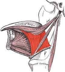
The laryngopharynx is located at the level of __. It extends from the upper border of the __ to the lower border of the __ cartillage. The _ is situated on each side of the larynx What are the muscles of the pharynx? |
-C3-C6 -epiglottis -cricoid -piriform -external layer (superior, middle, and inferior constrictors) and the longitudinal layer (palatopharyngeus, salpingopharyngeus and stylopharyngeus) |
|
|
The __ muscle is the landmark structure that separates the superior constrictors from the middle and inferior constrictor muscles All the muscles of the pharynx are supplied by the _ nerve with its pharyngeal branch plexus EXCEPT the _muscle which is innervated by the _ nerve What is the pharyngeal plexus formed by? What is the blood supply of the pharynx? |
-stylopharyngeus -CN 10 -stylopharyngeus -CN 9 -CN 10 (pharyngeal branch), CN 9 (afferent fibers), and sympathetic fibers from the superior cervical ganglion -ascending pharyngeal and inferior thyroid artery |
|
|
The _ connects the lower part of the pharynx with the trachea The larynx extends from __ where it becomes continuous with the trachea What are the functions of the the larynx? (3) |
larynx C3-C6 -to gaurd the air way passages, maintenance of a patient airway, and vocalization |
|
|
__ is at C4/C5. it has a laminae and a median elevation. (adams apple) __ cartilage at C6. It marks the end of the pharynx and larynx and the beginning of the esophagus and trachea _ cartilage is shaped like a vase for attachment of the arytenoid muscles _ cartilage is located in the ary-epiglottic fold __ cartilage is behind the root of the tongue and the body of the hyoid bone, and in front of the inlet of the larynx
|
thyroid cartilage cricoid cartilage arytenoid cartilage corniculate and cuneiform cartilages epiglottic cartilage |
|
|
__ joint is a synovial joint joint between the lamina of the cricoid cartilage and the inferior horn of the thyroid cartilage. it rotates the thyroid cartilage _ joint is synovial joint where the arytenoid cartilage will glide, rotate and rock at this joint |
cricothyroid joint cricoarytenoid joint |
|
|
Muscles of the larynx (extrinsic): _ and __ muscle will adduct the vocal cords. Which muscle adducts the vocal cords by bringing together the arytenoid cartilages? _ muscle will close the inlet of the larynx Intrinsic: _ is an adductor muscle *__ is the only abductor muscle* |
-transverse arytenoid and oblique arytenoid -oblique arytenoid -aryepiglottic -lateral cricoarytenoid -posterior cricoarytenoid
|
|
|
Muscles that regulate the vocal ligaments: |
thyroarytenoid muscle vocalis muscle cricothyroid muscle |
|
|
All the intrinsic muscles of the larynx are supplied by the __ nerve except the _ muscle which is innervated by the external laryngeal nerve from the superior laryngeal branch of the vagus nerve *damage to the recurrent laryngeal nerve may lead to __. Damage may occur secondary to a lung tumor, aortic aneurysm, trauma during thyroid surgery* |
recurrent laryngeal cricothyroid hoarseness |
|
|
*The sensory innervation of the larynx to the area above the vocal cords is provided by the _ nerve (superior laryngeal nerve). Below the vocal cords, the _- nerve supplies the mucosa with sensory innervation* What is the blood supply of the larynx?
|
-internal laryngeal nerve -recurrent laryngeal nerve -superior and inferior laryngeal artery |
|
|
The oral cavity: *posteriorly the oral pharynx communicates with the __ which is bounded by the palatoglossal arches laterally, the __ inferiorly and the ___ superiorly The upper lip drains into the _duct The __ palate is anterior 2/3 bony part the _ palate is the posterior 1/3 fibromuscular mobile part |
oropharynx epiglottis soft palate submandibular duct |
|
|
What is the hard palate formed by? The hard plate is covered by __ which forms a median raphe terminating in front of the incisive papilla The hard palate is ridge (__) which aids in the grabbing of food |
palatine process of the maxilla and the horizontal plates of the palatine bone -mucoperiostium -transverse palatine folds |
|
|
In the soft palates what two lateral folds are continuous with the uvula? What tonsils are located between those two folds? |
palatoglossal arch and palatopharyngeal arch -palatine tosils |
|
|
Muscles of the soft palate: Musculus Uvulae innervated by __ levator veli palatini innervated by__ Tensor veli palatini innervated by __ Palatoglossus muscle innervated by __ palatopharyngeus muscle innervated by __ |
vagus vagus manidbular (V3) vagus vagus |
|
|
What is the blood supply of the soft palate? All muscle the muscle of the soft palate are innervated by the vagus nerve except which muscle? what is that muscles innervation? Damage to the vagus nerve will cause the uvula to deviate to the _side damage to the glossopharyngeal (9) and vagus (10) nerve will result in __ loss of the gag reflex |
greater palatine artery from the descending palatine off the maxillary tensor veli palatini mandibualr (v3) opposite ipsilateral |
|
|
What are the main function of the tongue? What are the 4 lingual papillae? What is the space between the lateral and median glossoepiglottic folds? The pharyngeal portion of the mucous membrane forms what folds? (2) |
-taste, mastication, swallowing, and speech -filiform (conical), fungiform (taste buds), vallate (largest), and foliate (grooves and ridges) -epiglottic vallecula -lateral and medial glossoepiglottic folds |
|
|
All the extrinsic muscles of the tongue (genio-,hyo-, and styloglossus) are innervated by the __ nerve except the palatoglossus which is supplied by what nerve? *Damage to the CN 12 will produce __ atrophy of the tongue (wrinkled). when the tongue is protruded the tip of the tongue deviates towards the side of __* Blood supply of the tongue is the _ artery |
-vagus ipsilateral lesion lingual
|
|
|
*The anterior 2/3 of the tongue has general sensation supplied by the __ nerve, and taste by the __. The posterior 2/3 of tongue has general sensation from the __ nerve and taste from the __ nerve* |
lingual nerve (V3) chorda tympani (CN 7) CN 9 CN 9 |